Nickel in PDB 1e6v: Methyl-Coenzyme M Reductase From Methanopyrus Kandleri
Protein crystallography data
The structure of Methyl-Coenzyme M Reductase From Methanopyrus Kandleri, PDB code: 1e6v
was solved by
W.Grabarse,
U.Ermler,
with X-Ray Crystallography technique. A brief refinement statistics is given in the table below:
Resolution Low / High (Å)
|
30.00 /
2.70
|
Space group
|
P 21 21 21
|
Cell size a, b, c (Å), α, β, γ (°)
|
80.519,
115.740,
268.510,
90.00,
90.00,
90.00
|
R / Rfree (%)
|
23.9 /
27.8
|
Nickel Binding Sites:
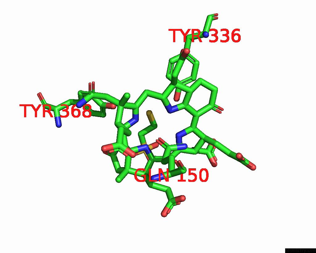
The binding sites of Nickel atom in the Methyl-Coenzyme M Reductase From Methanopyrus Kandleri
(pdb code 1e6v). This binding sites where shown within
5.0 Angstroms radius around Nickel atom.
In total 2 binding sites of Nickel where determined in the
Methyl-Coenzyme M Reductase From Methanopyrus Kandleri, PDB code: 1e6v:
Jump to Nickel binding site number:
1;
2;
Nickel binding site 1 out
of 2 in 1e6v
Go back to
Nickel Binding Sites List in 1e6v
Nickel binding site 1 out
of 2 in the Methyl-Coenzyme M Reductase From Methanopyrus Kandleri
Mono view
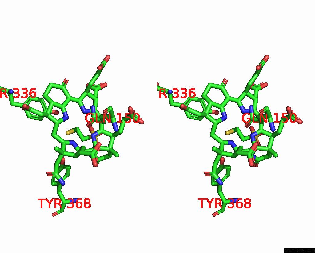
Stereo pair view
|
A full contact list of Nickel with other atoms in the Ni binding
site number 1 of Methyl-Coenzyme M Reductase From Methanopyrus Kandleri within 5.0Å range:
probe
|
atom
|
residue
|
distance (Å)
|
B
|
Occ
|
A:Ni1553
b:26.2
occ:1.00
|
NI
|
A:F431553
|
0.0
|
26.2
|
1.0
|
ND
|
A:F431553
|
2.0
|
26.1
|
1.0
|
NC
|
A:F431553
|
2.1
|
26.1
|
1.0
|
OE1
|
A:GLN150
|
2.1
|
19.0
|
1.0
|
NB
|
A:F431553
|
2.1
|
26.0
|
1.0
|
NA
|
A:F431553
|
2.1
|
26.1
|
1.0
|
S1
|
A:COM1555
|
2.5
|
17.3
|
1.0
|
C1A
|
A:F431553
|
2.9
|
26.2
|
1.0
|
C4B
|
A:F431553
|
2.9
|
25.9
|
1.0
|
C1C
|
A:F431553
|
3.0
|
26.1
|
1.0
|
CD
|
A:GLN150
|
3.0
|
18.6
|
1.0
|
C1D
|
A:F431553
|
3.1
|
26.0
|
1.0
|
C4D
|
A:F431553
|
3.1
|
26.2
|
1.0
|
C4C
|
A:F431553
|
3.1
|
26.1
|
1.0
|
C1B
|
A:F431553
|
3.2
|
25.9
|
1.0
|
C4A
|
A:F431553
|
3.3
|
26.0
|
1.0
|
CHA
|
A:F431553
|
3.3
|
26.1
|
1.0
|
CHC
|
A:F431553
|
3.3
|
26.0
|
1.0
|
C1
|
A:COM1555
|
3.4
|
17.0
|
1.0
|
CHB
|
A:F431553
|
3.4
|
25.9
|
1.0
|
NE2
|
A:GLN150
|
3.4
|
18.9
|
1.0
|
CHD
|
A:F431553
|
3.5
|
26.0
|
1.0
|
N5B
|
A:F431553
|
3.9
|
25.7
|
1.0
|
C2
|
A:COM1555
|
4.2
|
17.0
|
1.0
|
C3A
|
A:F431553
|
4.3
|
25.9
|
1.0
|
C3B
|
A:F431553
|
4.3
|
25.6
|
1.0
|
C3D
|
A:F431553
|
4.3
|
26.1
|
1.0
|
C2C
|
A:F431553
|
4.4
|
26.3
|
1.0
|
OH
|
D:TYR336
|
4.4
|
16.2
|
1.0
|
C2A
|
A:F431553
|
4.4
|
26.1
|
1.0
|
C2D
|
A:F431553
|
4.4
|
25.9
|
1.0
|
CG
|
A:GLN150
|
4.4
|
18.4
|
1.0
|
C3C
|
A:F431553
|
4.4
|
26.2
|
1.0
|
OH
|
E:TYR368
|
4.4
|
25.4
|
1.0
|
CAA
|
A:F431553
|
4.6
|
25.8
|
1.0
|
C2B
|
A:F431553
|
4.6
|
25.8
|
1.0
|
CAB
|
A:F431553
|
5.0
|
25.4
|
1.0
|
|
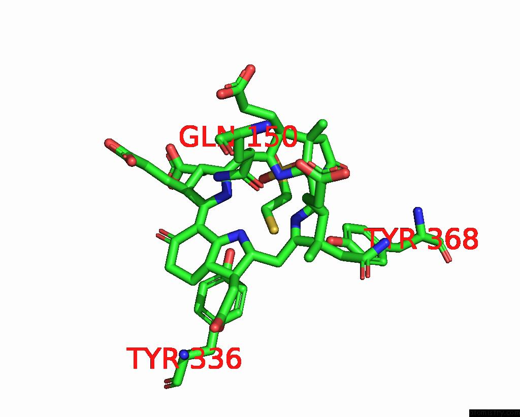
Nickel binding site 2 out
of 2 in 1e6v
Go back to
Nickel Binding Sites List in 1e6v
Nickel binding site 2 out
of 2 in the Methyl-Coenzyme M Reductase From Methanopyrus Kandleri
Mono view
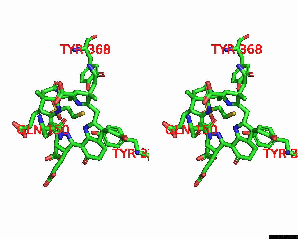
Stereo pair view
|
A full contact list of Nickel with other atoms in the Ni binding
site number 2 of Methyl-Coenzyme M Reductase From Methanopyrus Kandleri within 5.0Å range:
probe
|
atom
|
residue
|
distance (Å)
|
B
|
Occ
|
D:Ni1553
b:26.2
occ:1.00
|
NI
|
D:F431553
|
0.0
|
26.2
|
1.0
|
ND
|
D:F431553
|
2.0
|
26.1
|
1.0
|
NC
|
D:F431553
|
2.1
|
26.1
|
1.0
|
OE1
|
D:GLN150
|
2.1
|
19.0
|
1.0
|
NB
|
D:F431553
|
2.1
|
26.0
|
1.0
|
NA
|
D:F431553
|
2.1
|
26.1
|
1.0
|
S1
|
D:COM1555
|
2.5
|
17.3
|
1.0
|
C1A
|
D:F431553
|
2.9
|
26.2
|
1.0
|
C4B
|
D:F431553
|
2.9
|
25.9
|
1.0
|
C1C
|
D:F431553
|
3.0
|
26.1
|
1.0
|
CD
|
D:GLN150
|
3.0
|
18.6
|
1.0
|
C1D
|
D:F431553
|
3.1
|
26.0
|
1.0
|
C4D
|
D:F431553
|
3.1
|
26.2
|
1.0
|
C4C
|
D:F431553
|
3.1
|
26.1
|
1.0
|
C1B
|
D:F431553
|
3.2
|
25.9
|
1.0
|
C4A
|
D:F431553
|
3.3
|
26.0
|
1.0
|
CHA
|
D:F431553
|
3.3
|
26.1
|
1.0
|
CHC
|
D:F431553
|
3.3
|
26.0
|
1.0
|
C1
|
D:COM1555
|
3.4
|
17.0
|
1.0
|
CHB
|
D:F431553
|
3.4
|
25.9
|
1.0
|
NE2
|
D:GLN150
|
3.4
|
18.9
|
1.0
|
CHD
|
D:F431553
|
3.5
|
26.0
|
1.0
|
N5B
|
D:F431553
|
3.9
|
25.7
|
1.0
|
C2
|
D:COM1555
|
4.2
|
17.0
|
1.0
|
C3A
|
D:F431553
|
4.3
|
25.9
|
1.0
|
C3D
|
D:F431553
|
4.3
|
26.1
|
1.0
|
C3B
|
D:F431553
|
4.3
|
25.6
|
1.0
|
C2C
|
D:F431553
|
4.4
|
26.3
|
1.0
|
C2A
|
D:F431553
|
4.4
|
26.1
|
1.0
|
OH
|
A:TYR336
|
4.4
|
16.2
|
1.0
|
C2D
|
D:F431553
|
4.4
|
25.9
|
1.0
|
CG
|
D:GLN150
|
4.4
|
18.4
|
1.0
|
C3C
|
D:F431553
|
4.4
|
26.2
|
1.0
|
OH
|
B:TYR368
|
4.4
|
25.4
|
1.0
|
CAA
|
D:F431553
|
4.6
|
25.8
|
1.0
|
C2B
|
D:F431553
|
4.6
|
25.8
|
1.0
|
CAB
|
D:F431553
|
5.0
|
25.4
|
1.0
|
|
Reference:
W.Grabarse,
F.Mahlert,
S.Shima,
R.K.Thauer,
U.Ermler.
Comparison of Three Methyl-Coenzyme M Reductases From Phylogenetically Distant Organisms: Unusual Amino Acid Modification, Conservation and Adaptation J.Mol.Biol. V. 303 329 2000.
ISSN: ISSN 0022-2836
PubMed: 11023796
DOI: 10.1006/JMBI.2000.4136
Page generated: Wed Oct 9 14:47:51 2024
|