Nickel »
PDB 1fwc-1j6p »
1g2a »
Nickel in PDB 1g2a: The Crystal Structure of E.Coli Peptide Deformylase Complexed with Actinonin
Enzymatic activity of The Crystal Structure of E.Coli Peptide Deformylase Complexed with Actinonin
All present enzymatic activity of The Crystal Structure of E.Coli Peptide Deformylase Complexed with Actinonin:
3.5.1.31;
3.5.1.31;
Protein crystallography data
The structure of The Crystal Structure of E.Coli Peptide Deformylase Complexed with Actinonin, PDB code: 1g2a
was solved by
J.M.Clements,
P.Beckett,
A.Brown,
C.Catlin,
M.Lobell,
S.Palan,
W.Thomas,
M.Whittaker,
P.J.Baker,
H.F.Rodgers,
V.Barynin,
D.W.Rice,
M.G.Hunter,
with X-Ray Crystallography technique. A brief refinement statistics is given in the table below:
| Resolution Low / High (Å) | 14.00 / 1.75 |
| Space group | C 1 2 1 |
| Cell size a, b, c (Å), α, β, γ (°) | 138.700, 63.100, 85.600, 90.00, 121.40, 90.00 |
| R / Rfree (%) | 19 / 25 |
Nickel Binding Sites:
The binding sites of Nickel atom in the The Crystal Structure of E.Coli Peptide Deformylase Complexed with Actinonin
(pdb code 1g2a). This binding sites where shown within
5.0 Angstroms radius around Nickel atom.
In total 3 binding sites of Nickel where determined in the The Crystal Structure of E.Coli Peptide Deformylase Complexed with Actinonin, PDB code: 1g2a:
Jump to Nickel binding site number: 1; 2; 3;
In total 3 binding sites of Nickel where determined in the The Crystal Structure of E.Coli Peptide Deformylase Complexed with Actinonin, PDB code: 1g2a:
Jump to Nickel binding site number: 1; 2; 3;
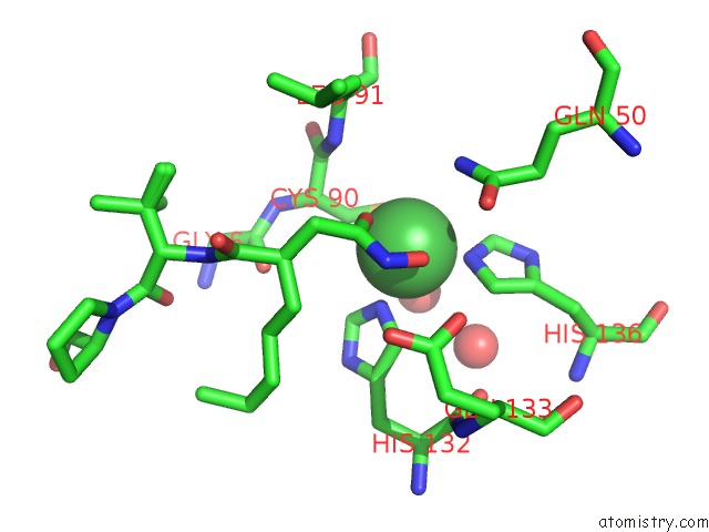
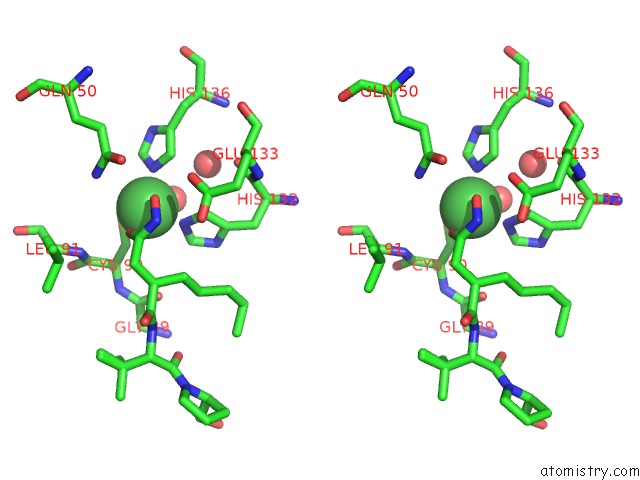
Nickel binding site 1 out of 3 in 1g2a
Go back to
Nickel binding site 1 out
of 3 in the The Crystal Structure of E.Coli Peptide Deformylase Complexed with Actinonin
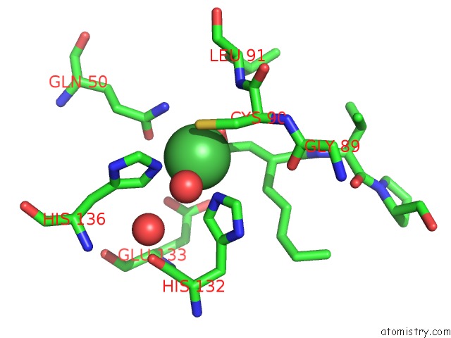
Mono view
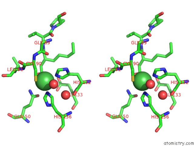
Stereo pair view
Mono view
Stereo pair view
A full contact list of Nickel with other atoms in the Ni binding
site number 1 of The Crystal Structure of E.Coli Peptide Deformylase Complexed with Actinonin within 5.0Å range:
|
Nickel binding site 2 out of 3 in 1g2a
Go back to
Nickel binding site 2 out
of 3 in the The Crystal Structure of E.Coli Peptide Deformylase Complexed with Actinonin
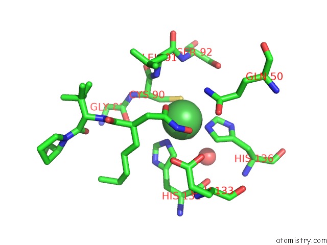
Mono view
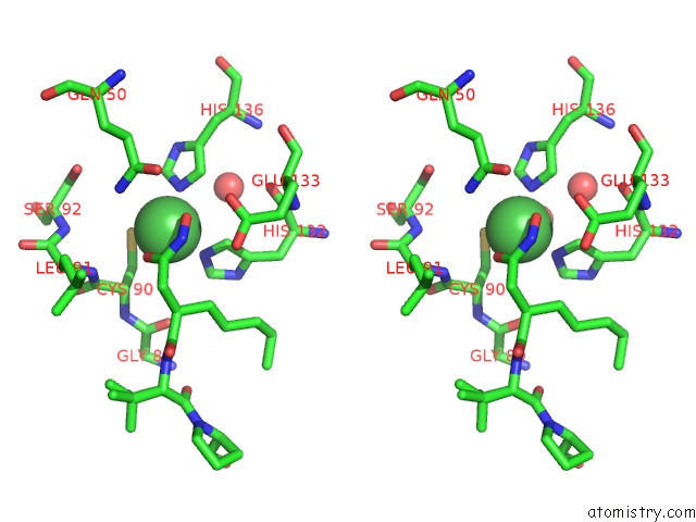
Stereo pair view
Mono view
Stereo pair view
A full contact list of Nickel with other atoms in the Ni binding
site number 2 of The Crystal Structure of E.Coli Peptide Deformylase Complexed with Actinonin within 5.0Å range:
|
Nickel binding site 3 out of 3 in 1g2a
Go back to
Nickel binding site 3 out
of 3 in the The Crystal Structure of E.Coli Peptide Deformylase Complexed with Actinonin
Mono view
Stereo pair view
Mono view
Stereo pair view
A full contact list of Nickel with other atoms in the Ni binding
site number 3 of The Crystal Structure of E.Coli Peptide Deformylase Complexed with Actinonin within 5.0Å range:
|
Reference:
J.M.Clements,
R.P.Beckett,
A.Brown,
G.Catlin,
M.Lobell,
S.Palan,
W.Thomas,
M.Whittaker,
S.Wood,
S.Salama,
P.J.Baker,
H.F.Rodgers,
V.Barynin,
D.W.Rice,
M.G.Hunter.
Antibiotic Activity and Characterization of Bb-3497, A Novel Peptide Deformylase Inhibitor. Antimicrob.Agents Chemother. V. 45 563 2001.
ISSN: ISSN 0066-4804
PubMed: 11158755
DOI: 10.1128/AAC.45.2.563-570.2001
Page generated: Wed Oct 9 14:54:36 2024
ISSN: ISSN 0066-4804
PubMed: 11158755
DOI: 10.1128/AAC.45.2.563-570.2001
Last articles
Zn in 9MJ5Zn in 9HNW
Zn in 9G0L
Zn in 9FNE
Zn in 9DZN
Zn in 9E0I
Zn in 9D32
Zn in 9DAK
Zn in 8ZXC
Zn in 8ZUF