Nickel »
PDB 1fwc-1j6p »
1h2a »
Nickel in PDB 1h2a: Single Crystals of Hydrogenase From Desulfovibrio Vulgaris
Enzymatic activity of Single Crystals of Hydrogenase From Desulfovibrio Vulgaris
All present enzymatic activity of Single Crystals of Hydrogenase From Desulfovibrio Vulgaris:
1.18.99.1;
1.18.99.1;
Protein crystallography data
The structure of Single Crystals of Hydrogenase From Desulfovibrio Vulgaris, PDB code: 1h2a
was solved by
Y.Higuchi,
N.Yasuoka,
with X-Ray Crystallography technique. A brief refinement statistics is given in the table below:
| Resolution Low / High (Å) | 20.00 / 1.80 |
| Space group | P 21 21 21 |
| Cell size a, b, c (Å), α, β, γ (°) | 101.500, 126.500, 66.510, 90.00, 90.00, 90.00 |
| R / Rfree (%) | 22.9 / 27.8 |
Other elements in 1h2a:
The structure of Single Crystals of Hydrogenase From Desulfovibrio Vulgaris also contains other interesting chemical elements:
| Magnesium | (Mg) | 1 atom |
| Iron | (Fe) | 12 atoms |
Nickel Binding Sites:
The binding sites of Nickel atom in the Single Crystals of Hydrogenase From Desulfovibrio Vulgaris
(pdb code 1h2a). This binding sites where shown within
5.0 Angstroms radius around Nickel atom.
In total only one binding site of Nickel was determined in the Single Crystals of Hydrogenase From Desulfovibrio Vulgaris, PDB code: 1h2a:
In total only one binding site of Nickel was determined in the Single Crystals of Hydrogenase From Desulfovibrio Vulgaris, PDB code: 1h2a:
Nickel binding site 1 out of 1 in 1h2a
Go back to
Nickel binding site 1 out
of 1 in the Single Crystals of Hydrogenase From Desulfovibrio Vulgaris
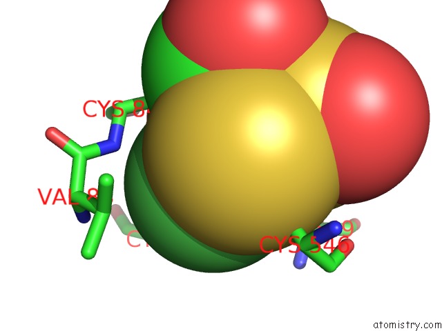
Mono view
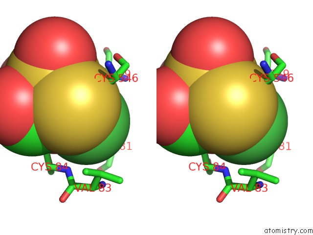
Stereo pair view
Mono view
Stereo pair view
A full contact list of Nickel with other atoms in the Ni binding
site number 1 of Single Crystals of Hydrogenase From Desulfovibrio Vulgaris within 5.0Å range:
|
Reference:
Y.Higuchi,
T.Yagi,
N.Yasuoka.
Unusual Ligand Structure in Ni-Fe Active Center and An Additional Mg Site in Hydrogenase Revealed By High Resolution X-Ray Structure Analysis. Structure V. 5 1671 1997.
ISSN: ISSN 0969-2126
PubMed: 9438867
DOI: 10.1016/S0969-2126(97)00313-4
Page generated: Wed Oct 9 14:54:44 2024
ISSN: ISSN 0969-2126
PubMed: 9438867
DOI: 10.1016/S0969-2126(97)00313-4
Last articles
Zn in 9J0NZn in 9J0O
Zn in 9J0P
Zn in 9FJX
Zn in 9EKB
Zn in 9C0F
Zn in 9CAH
Zn in 9CH0
Zn in 9CH3
Zn in 9CH1