Nickel »
PDB 1fwc-1j6p »
1hbm »
Nickel in PDB 1hbm: Methyl-Coenzyme M Reductase Enzyme Product Complex
Protein crystallography data
The structure of Methyl-Coenzyme M Reductase Enzyme Product Complex, PDB code: 1hbm
was solved by
U.Ermler,
W.Grabarse,
with X-Ray Crystallography technique. A brief refinement statistics is given in the table below:
| Resolution Low / High (Å) | 10.00 / 1.80 |
| Space group | P 1 21 1 |
| Cell size a, b, c (Å), α, β, γ (°) | 82.000, 118.300, 122.800, 90.00, 92.00, 90.00 |
| R / Rfree (%) | 16.4 / 19.3 |
Other elements in 1hbm:
The structure of Methyl-Coenzyme M Reductase Enzyme Product Complex also contains other interesting chemical elements:
| Magnesium | (Mg) | 5 atoms |
| Zinc | (Zn) | 1 atom |
| Chlorine | (Cl) | 2 atoms |
| Sodium | (Na) | 9 atoms |
Nickel Binding Sites:
The binding sites of Nickel atom in the Methyl-Coenzyme M Reductase Enzyme Product Complex
(pdb code 1hbm). This binding sites where shown within
5.0 Angstroms radius around Nickel atom.
In total 2 binding sites of Nickel where determined in the Methyl-Coenzyme M Reductase Enzyme Product Complex, PDB code: 1hbm:
Jump to Nickel binding site number: 1; 2;
In total 2 binding sites of Nickel where determined in the Methyl-Coenzyme M Reductase Enzyme Product Complex, PDB code: 1hbm:
Jump to Nickel binding site number: 1; 2;
Nickel binding site 1 out of 2 in 1hbm
Go back to
Nickel binding site 1 out
of 2 in the Methyl-Coenzyme M Reductase Enzyme Product Complex
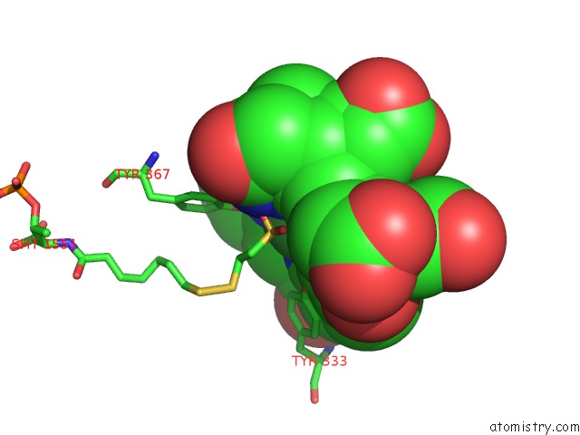
Mono view
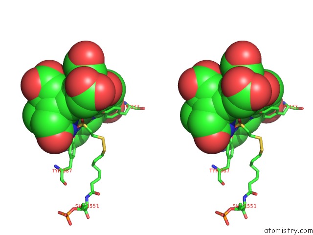
Stereo pair view
Mono view
Stereo pair view
A full contact list of Nickel with other atoms in the Ni binding
site number 1 of Methyl-Coenzyme M Reductase Enzyme Product Complex within 5.0Å range:
|
Nickel binding site 2 out of 2 in 1hbm
Go back to
Nickel binding site 2 out
of 2 in the Methyl-Coenzyme M Reductase Enzyme Product Complex
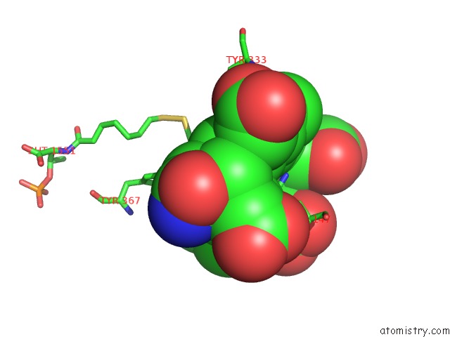
Mono view
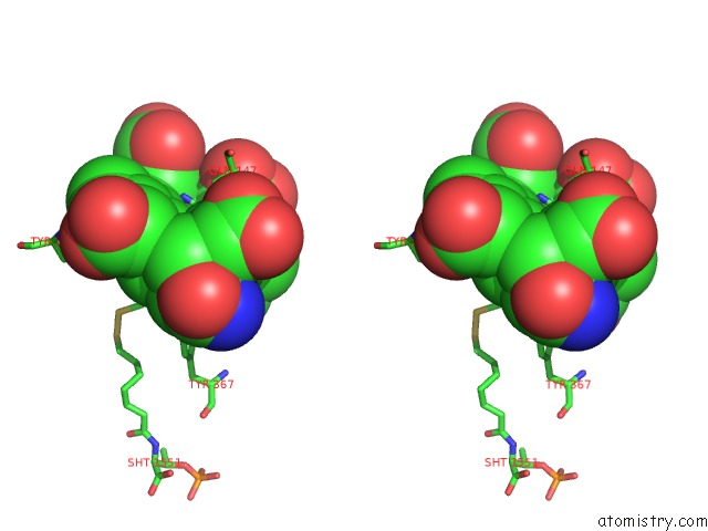
Stereo pair view
Mono view
Stereo pair view
A full contact list of Nickel with other atoms in the Ni binding
site number 2 of Methyl-Coenzyme M Reductase Enzyme Product Complex within 5.0Å range:
|
Reference:
W.Grabarse,
F.Mahlert,
E.C.Duin,
M.Goubeaud,
S.Shima,
R.K.Thauer,
V.Lamzin,
U.Ermler.
On the Mechanism of Biological Methane Formation: Structural Evidence For Conformational Changes in Methyl-Coenzyme M Reductase Upon Substrate Binding J.Mol.Biol. V. 309 315 2001.
ISSN: ISSN 0022-2836
PubMed: 11491299
DOI: 10.1006/JMBI.2001.4647
Page generated: Wed Oct 9 14:55:30 2024
ISSN: ISSN 0022-2836
PubMed: 11491299
DOI: 10.1006/JMBI.2001.4647
Last articles
Zn in 9MJ5Zn in 9HNW
Zn in 9G0L
Zn in 9FNE
Zn in 9DZN
Zn in 9E0I
Zn in 9D32
Zn in 9DAK
Zn in 8ZXC
Zn in 8ZUF