Nickel »
PDB 1t6u-1xmk »
1xmk »
Nickel in PDB 1xmk: The Crystal Structure of the Zb Domain From the Rna Editing Enzyme ADAR1
Protein crystallography data
The structure of The Crystal Structure of the Zb Domain From the Rna Editing Enzyme ADAR1, PDB code: 1xmk
was solved by
A.Athanasiadis,
D.Placido,
S.Maas,
B.A.Brown Ii,
K.Lowenhaupt,
A.Rich,
with X-Ray Crystallography technique. A brief refinement statistics is given in the table below:
| Resolution Low / High (Å) | 10.00 / 0.97 |
| Space group | P 21 21 21 |
| Cell size a, b, c (Å), α, β, γ (°) | 35.557, 43.526, 45.471, 90.00, 90.00, 90.00 |
| R / Rfree (%) | 14.5 / 18.3 |
Other elements in 1xmk:
The structure of The Crystal Structure of the Zb Domain From the Rna Editing Enzyme ADAR1 also contains other interesting chemical elements:
| Cadmium | (Cd) | 2 atoms |
| Chlorine | (Cl) | 2 atoms |
Nickel Binding Sites:
The binding sites of Nickel atom in the The Crystal Structure of the Zb Domain From the Rna Editing Enzyme ADAR1
(pdb code 1xmk). This binding sites where shown within
5.0 Angstroms radius around Nickel atom.
In total only one binding site of Nickel was determined in the The Crystal Structure of the Zb Domain From the Rna Editing Enzyme ADAR1, PDB code: 1xmk:
In total only one binding site of Nickel was determined in the The Crystal Structure of the Zb Domain From the Rna Editing Enzyme ADAR1, PDB code: 1xmk:
Nickel binding site 1 out of 1 in 1xmk
Go back to
Nickel binding site 1 out
of 1 in the The Crystal Structure of the Zb Domain From the Rna Editing Enzyme ADAR1
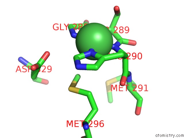
Mono view
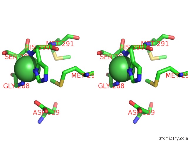
Stereo pair view
Mono view
Stereo pair view
A full contact list of Nickel with other atoms in the Ni binding
site number 1 of The Crystal Structure of the Zb Domain From the Rna Editing Enzyme ADAR1 within 5.0Å range:
|
Reference:
A.Athanasiadis,
D.Placido,
S.Maas,
B.A.Brown Ii,
K.Lowenhaupt,
A.Rich.
The Crystal Structure of the Z[Beta] Domain of the Rna-Editing Enzyme ADAR1 Reveals Distinct Conserved Surfaces Among Z-Domains. J.Mol.Biol. V. 351 496 2005.
ISSN: ISSN 0022-2836
PubMed: 16023667
DOI: 10.1016/J.JMB.2005.06.028
Page generated: Wed Oct 9 16:31:53 2024
ISSN: ISSN 0022-2836
PubMed: 16023667
DOI: 10.1016/J.JMB.2005.06.028
Last articles
Zn in 9MJ5Zn in 9HNW
Zn in 9G0L
Zn in 9FNE
Zn in 9DZN
Zn in 9E0I
Zn in 9D32
Zn in 9DAK
Zn in 8ZXC
Zn in 8ZUF