Nickel »
PDB 1xu1-2c0n »
2bmr »
Nickel in PDB 2bmr: The Crystal Structure of Nitrobenzene Dioxygenase in Complex with 3- Nitrotoluene
Protein crystallography data
The structure of The Crystal Structure of Nitrobenzene Dioxygenase in Complex with 3- Nitrotoluene, PDB code: 2bmr
was solved by
R.Friemann,
M.M.Ivkovic-Jensen,
D.J.Lessner,
C.Yu,
D.T.Gibson,
R.E.Parales,
H.Eklund,
S.Ramaswamy,
with X-Ray Crystallography technique. A brief refinement statistics is given in the table below:
| Resolution Low / High (Å) | 52.55 / 1.50 |
| Space group | P 63 |
| Cell size a, b, c (Å), α, β, γ (°) | 121.625, 121.625, 84.002, 90.00, 90.00, 120.00 |
| R / Rfree (%) | 16.9 / 19.1 |
Other elements in 2bmr:
The structure of The Crystal Structure of Nitrobenzene Dioxygenase in Complex with 3- Nitrotoluene also contains other interesting chemical elements:
| Iron | (Fe) | 3 atoms |
Nickel Binding Sites:
The binding sites of Nickel atom in the The Crystal Structure of Nitrobenzene Dioxygenase in Complex with 3- Nitrotoluene
(pdb code 2bmr). This binding sites where shown within
5.0 Angstroms radius around Nickel atom.
In total 2 binding sites of Nickel where determined in the The Crystal Structure of Nitrobenzene Dioxygenase in Complex with 3- Nitrotoluene, PDB code: 2bmr:
Jump to Nickel binding site number: 1; 2;
In total 2 binding sites of Nickel where determined in the The Crystal Structure of Nitrobenzene Dioxygenase in Complex with 3- Nitrotoluene, PDB code: 2bmr:
Jump to Nickel binding site number: 1; 2;
Nickel binding site 1 out of 2 in 2bmr
Go back to
Nickel binding site 1 out
of 2 in the The Crystal Structure of Nitrobenzene Dioxygenase in Complex with 3- Nitrotoluene
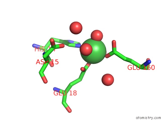
Mono view
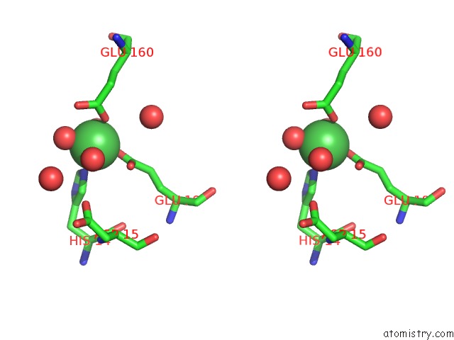
Stereo pair view
Mono view
Stereo pair view
A full contact list of Nickel with other atoms in the Ni binding
site number 1 of The Crystal Structure of Nitrobenzene Dioxygenase in Complex with 3- Nitrotoluene within 5.0Å range:
|
Nickel binding site 2 out of 2 in 2bmr
Go back to
Nickel binding site 2 out
of 2 in the The Crystal Structure of Nitrobenzene Dioxygenase in Complex with 3- Nitrotoluene
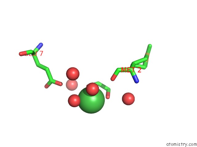
Mono view
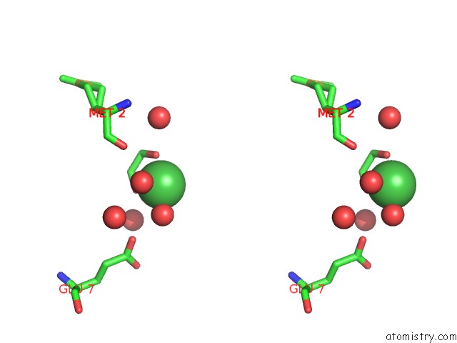
Stereo pair view
Mono view
Stereo pair view
A full contact list of Nickel with other atoms in the Ni binding
site number 2 of The Crystal Structure of Nitrobenzene Dioxygenase in Complex with 3- Nitrotoluene within 5.0Å range:
|
Reference:
R.Friemann,
M.M.Ivkovic-Jensen,
D.J.Lessner,
C.L.Yu,
D.T.Gibson,
R.E.Parales,
H.Eklund,
S.Ramaswamy.
Structural Insight Into the Dioxygenation of Nitroarene Compounds: the Crystal Structure of Nitrobenzene Dioxygenase. J. Mol. Biol. V. 348 1139 2005.
ISSN: ISSN 0022-2836
PubMed: 15854650
DOI: 10.1016/J.JMB.2005.03.052
Page generated: Wed Oct 9 16:39:37 2024
ISSN: ISSN 0022-2836
PubMed: 15854650
DOI: 10.1016/J.JMB.2005.03.052
Last articles
Zn in 9J0NZn in 9J0O
Zn in 9J0P
Zn in 9FJX
Zn in 9EKB
Zn in 9C0F
Zn in 9CAH
Zn in 9CH0
Zn in 9CH3
Zn in 9CH1