Nickel »
PDB 2c21-2gw3 »
2fcp »
Nickel in PDB 2fcp: Ferric Hydroxamate Uptake Receptor (Fhua) From E.Coli
Protein crystallography data
The structure of Ferric Hydroxamate Uptake Receptor (Fhua) From E.Coli, PDB code: 2fcp
was solved by
E.Hofmann,
A.D.Ferguson,
K.Diederichs,
W.Welte,
with X-Ray Crystallography technique. A brief refinement statistics is given in the table below:
| Resolution Low / High (Å) | 30.00 / 2.50 |
| Space group | P 61 |
| Cell size a, b, c (Å), α, β, γ (°) | 171.550, 171.550, 87.650, 90.00, 90.00, 120.00 |
| R / Rfree (%) | 24.2 / 28.3 |
Nickel Binding Sites:
The binding sites of Nickel atom in the Ferric Hydroxamate Uptake Receptor (Fhua) From E.Coli
(pdb code 2fcp). This binding sites where shown within
5.0 Angstroms radius around Nickel atom.
In total 2 binding sites of Nickel where determined in the Ferric Hydroxamate Uptake Receptor (Fhua) From E.Coli, PDB code: 2fcp:
Jump to Nickel binding site number: 1; 2;
In total 2 binding sites of Nickel where determined in the Ferric Hydroxamate Uptake Receptor (Fhua) From E.Coli, PDB code: 2fcp:
Jump to Nickel binding site number: 1; 2;
Nickel binding site 1 out of 2 in 2fcp
Go back to
Nickel binding site 1 out
of 2 in the Ferric Hydroxamate Uptake Receptor (Fhua) From E.Coli
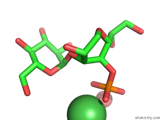
Mono view
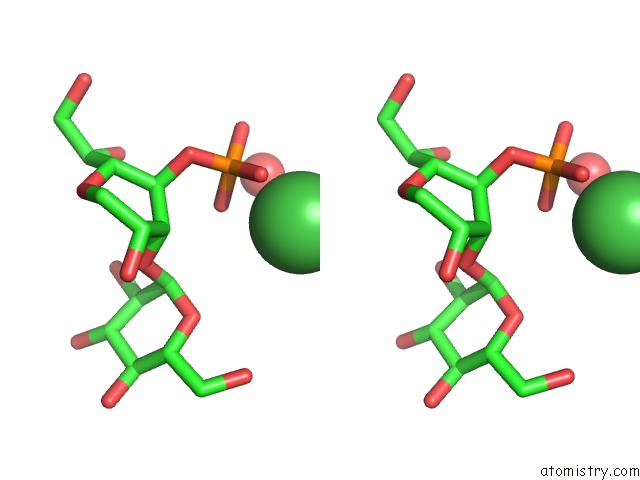
Stereo pair view
Mono view
Stereo pair view
A full contact list of Nickel with other atoms in the Ni binding
site number 1 of Ferric Hydroxamate Uptake Receptor (Fhua) From E.Coli within 5.0Å range:
|
Nickel binding site 2 out of 2 in 2fcp
Go back to
Nickel binding site 2 out
of 2 in the Ferric Hydroxamate Uptake Receptor (Fhua) From E.Coli
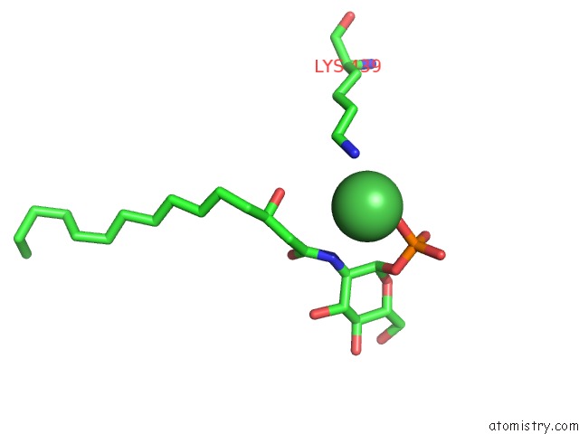
Mono view
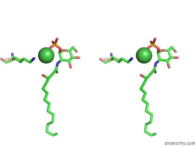
Stereo pair view
Mono view
Stereo pair view
A full contact list of Nickel with other atoms in the Ni binding
site number 2 of Ferric Hydroxamate Uptake Receptor (Fhua) From E.Coli within 5.0Å range:
|
Reference:
A.D.Ferguson,
E.Hofmann,
J.W.Coulton,
K.Diederichs,
W.Welte.
Siderophore-Mediated Iron Transport: Crystal Structure of Fhua with Bound Lipopolysaccharide. Science V. 282 2215 1998.
ISSN: ISSN 0036-8075
PubMed: 9856937
DOI: 10.1126/SCIENCE.282.5397.2215
Page generated: Wed Oct 9 16:42:59 2024
ISSN: ISSN 0036-8075
PubMed: 9856937
DOI: 10.1126/SCIENCE.282.5397.2215
Last articles
Zn in 9J0NZn in 9J0O
Zn in 9J0P
Zn in 9FJX
Zn in 9EKB
Zn in 9C0F
Zn in 9CAH
Zn in 9CH0
Zn in 9CH3
Zn in 9CH1