Nickel »
PDB 2c21-2gw3 »
2glz »
Nickel in PDB 2glz: Crystal Structure of A Formylmethanofuran Dehydrogenase Subunit E-Like Protein (DHAF_2992) From Desulfitobacterium Hafniense Dcb-2 at 1.45 A Resolution
Protein crystallography data
The structure of Crystal Structure of A Formylmethanofuran Dehydrogenase Subunit E-Like Protein (DHAF_2992) From Desulfitobacterium Hafniense Dcb-2 at 1.45 A Resolution, PDB code: 2glz
was solved by
Joint Center For Structural Genomics (Jcsg),
with X-Ray Crystallography technique. A brief refinement statistics is given in the table below:
| Resolution Low / High (Å) | 27.20 / 1.45 |
| Space group | P 21 21 21 |
| Cell size a, b, c (Å), α, β, γ (°) | 46.425, 84.791, 100.711, 90.00, 90.00, 90.00 |
| R / Rfree (%) | 17.1 / 19.8 |
Other elements in 2glz:
The structure of Crystal Structure of A Formylmethanofuran Dehydrogenase Subunit E-Like Protein (DHAF_2992) From Desulfitobacterium Hafniense Dcb-2 at 1.45 A Resolution also contains other interesting chemical elements:
| Zinc | (Zn) | 2 atoms |
Nickel Binding Sites:
The binding sites of Nickel atom in the Crystal Structure of A Formylmethanofuran Dehydrogenase Subunit E-Like Protein (DHAF_2992) From Desulfitobacterium Hafniense Dcb-2 at 1.45 A Resolution
(pdb code 2glz). This binding sites where shown within
5.0 Angstroms radius around Nickel atom.
In total 2 binding sites of Nickel where determined in the Crystal Structure of A Formylmethanofuran Dehydrogenase Subunit E-Like Protein (DHAF_2992) From Desulfitobacterium Hafniense Dcb-2 at 1.45 A Resolution, PDB code: 2glz:
Jump to Nickel binding site number: 1; 2;
In total 2 binding sites of Nickel where determined in the Crystal Structure of A Formylmethanofuran Dehydrogenase Subunit E-Like Protein (DHAF_2992) From Desulfitobacterium Hafniense Dcb-2 at 1.45 A Resolution, PDB code: 2glz:
Jump to Nickel binding site number: 1; 2;
Nickel binding site 1 out of 2 in 2glz
Go back to
Nickel binding site 1 out
of 2 in the Crystal Structure of A Formylmethanofuran Dehydrogenase Subunit E-Like Protein (DHAF_2992) From Desulfitobacterium Hafniense Dcb-2 at 1.45 A Resolution
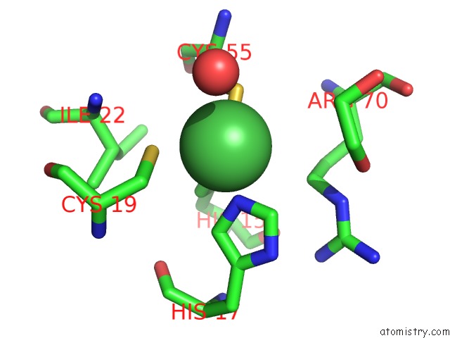
Mono view
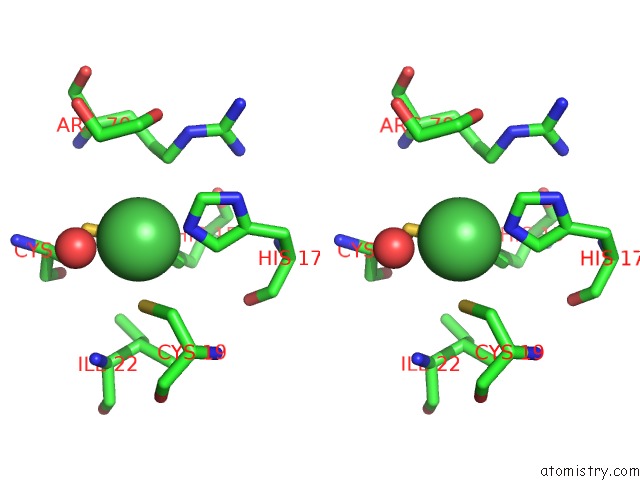
Stereo pair view
Mono view
Stereo pair view
A full contact list of Nickel with other atoms in the Ni binding
site number 1 of Crystal Structure of A Formylmethanofuran Dehydrogenase Subunit E-Like Protein (DHAF_2992) From Desulfitobacterium Hafniense Dcb-2 at 1.45 A Resolution within 5.0Å range:
|
Nickel binding site 2 out of 2 in 2glz
Go back to
Nickel binding site 2 out
of 2 in the Crystal Structure of A Formylmethanofuran Dehydrogenase Subunit E-Like Protein (DHAF_2992) From Desulfitobacterium Hafniense Dcb-2 at 1.45 A Resolution
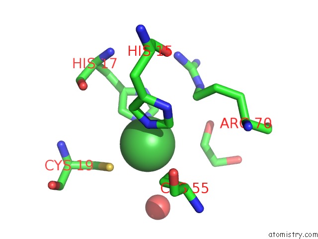
Mono view
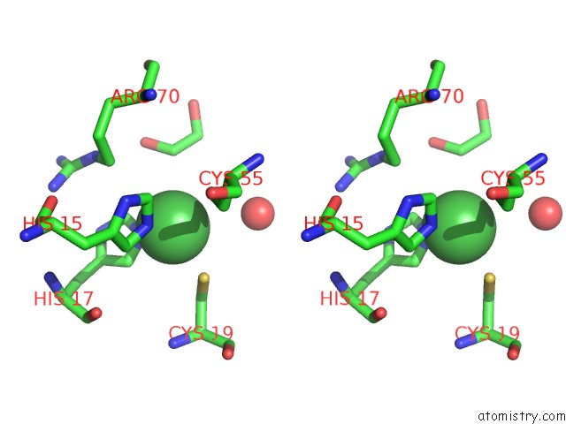
Stereo pair view
Mono view
Stereo pair view
A full contact list of Nickel with other atoms in the Ni binding
site number 2 of Crystal Structure of A Formylmethanofuran Dehydrogenase Subunit E-Like Protein (DHAF_2992) From Desulfitobacterium Hafniense Dcb-2 at 1.45 A Resolution within 5.0Å range:
|
Reference:
H.L.Axelrod,
D.Das,
P.Abdubek,
T.Astakhova,
C.Bakolitsa,
D.Carlton,
C.Chen,
H.J.Chiu,
T.Clayton,
M.C.Deller,
L.Duan,
K.Ellrott,
C.L.Farr,
J.Feuerhelm,
J.C.Grant,
A.Grzechnik,
G.W.Han,
L.Jaroszewski,
K.K.Jin,
H.E.Klock,
M.W.Knuth,
P.Kozbial,
S.S.Krishna,
A.Kumar,
W.W.Lam,
D.Marciano,
D.Mcmullan,
M.D.Miller,
A.T.Morse,
E.Nigoghossian,
A.Nopakun,
L.Okach,
C.Puckett,
R.Reyes,
N.Sefcovic,
H.J.Tien,
C.B.Trame,
H.Van Den Bedem,
D.Weekes,
T.Wooten,
Q.Xu,
K.O.Hodgson,
J.Wooley,
M.A.Elsliger,
A.M.Deacon,
A.Godzik,
S.A.Lesley,
I.A.Wilson.
Structures of Three Members of Pfam PF02663 (Fmde) Implicated in Microbial Methanogenesis Reveal A Conserved Alpha+Beta Core Domain and An Auxiliary C-Terminal Treble-Clef Zinc Finger. Acta Crystallogr.,Sect.F V. 66 1335 2010.
ISSN: ESSN 1744-3091
PubMed: 20944230
DOI: 10.1107/S1744309110020166
Page generated: Wed Oct 9 16:44:13 2024
ISSN: ESSN 1744-3091
PubMed: 20944230
DOI: 10.1107/S1744309110020166
Last articles
Zn in 9J0NZn in 9J0O
Zn in 9J0P
Zn in 9FJX
Zn in 9EKB
Zn in 9C0F
Zn in 9CAH
Zn in 9CH0
Zn in 9CH3
Zn in 9CH1