Nickel »
PDB 2gw4-2pos »
2isz »
Nickel in PDB 2isz: Crystal Structure of A Two-Domain Ider-Dna Complex Crystal Form I
Protein crystallography data
The structure of Crystal Structure of A Two-Domain Ider-Dna Complex Crystal Form I, PDB code: 2isz
was solved by
G.Wisedchaisri,
C.J.Chou,
M.Wu,
C.Roach,
A.E.Rice,
R.K.Holmes,
C.Beeson,
W.G.Hol,
with X-Ray Crystallography technique. A brief refinement statistics is given in the table below:
| Resolution Low / High (Å) | 50.00 / 2.40 |
| Space group | P 1 |
| Cell size a, b, c (Å), α, β, γ (°) | 53.832, 69.739, 76.461, 106.57, 104.85, 99.66 |
| R / Rfree (%) | 19.9 / 23.9 |
Other elements in 2isz:
The structure of Crystal Structure of A Two-Domain Ider-Dna Complex Crystal Form I also contains other interesting chemical elements:
| Sodium | (Na) | 3 atoms |
Nickel Binding Sites:
Pages:
>>> Page 1 <<< Page 2, Binding sites: 11 - 12;Binding sites:
The binding sites of Nickel atom in the Crystal Structure of A Two-Domain Ider-Dna Complex Crystal Form I (pdb code 2isz). This binding sites where shown within 5.0 Angstroms radius around Nickel atom.In total 12 binding sites of Nickel where determined in the Crystal Structure of A Two-Domain Ider-Dna Complex Crystal Form I, PDB code: 2isz:
Jump to Nickel binding site number: 1; 2; 3; 4; 5; 6; 7; 8; 9; 10;
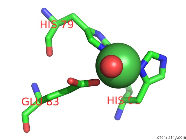
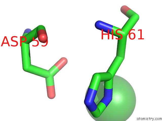
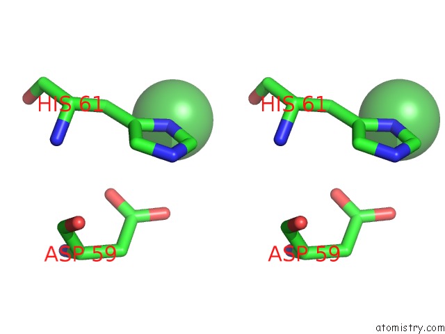
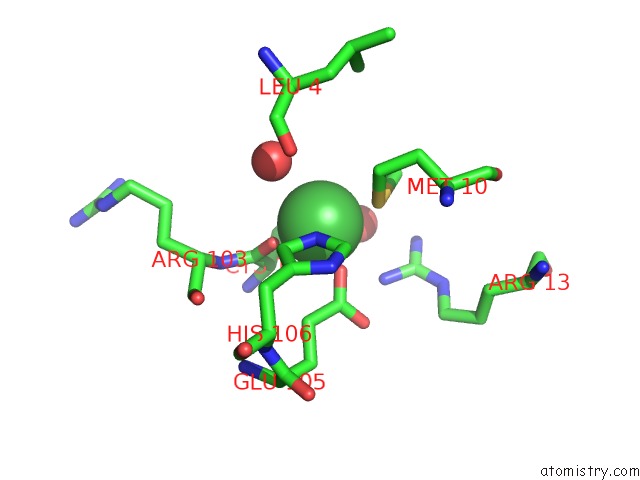
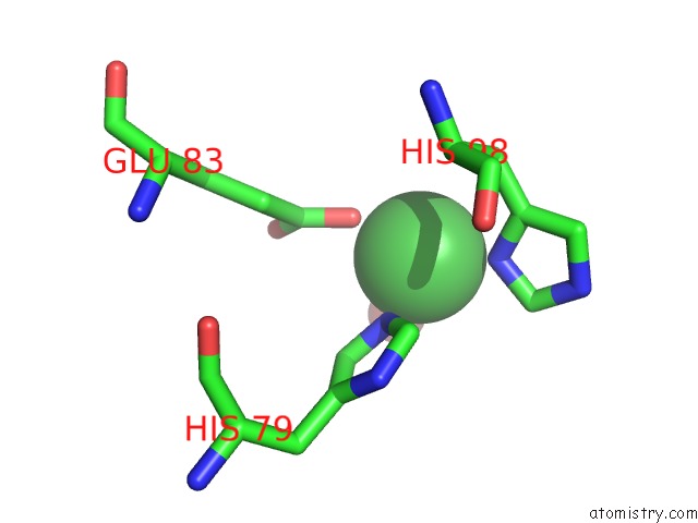
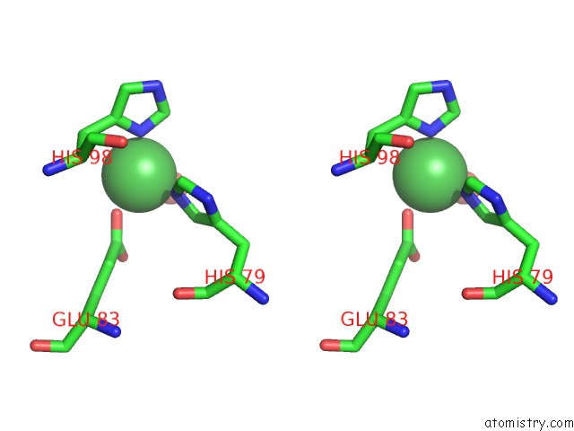
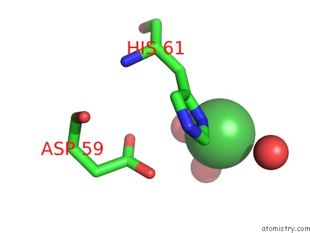
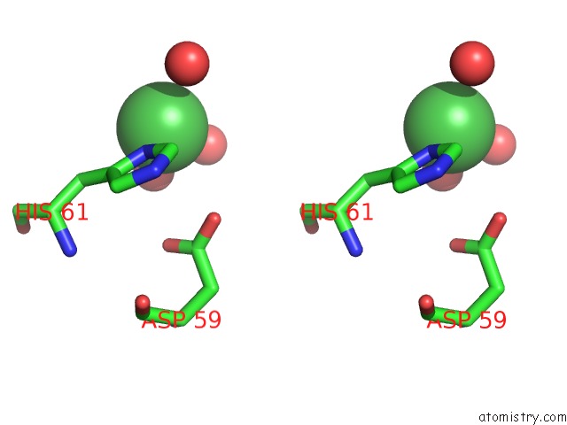
Nickel binding site 1 out of 12 in 2isz
Go back to
Nickel binding site 1 out
of 12 in the Crystal Structure of A Two-Domain Ider-Dna Complex Crystal Form I
Mono view
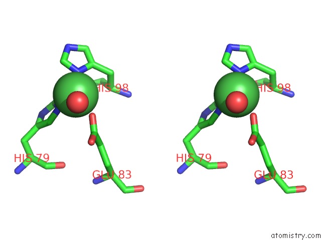
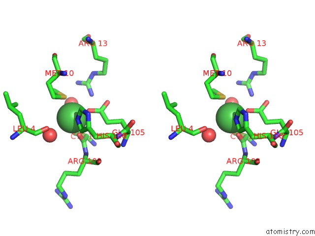
Stereo pair view
Mono view
Stereo pair view
A full contact list of Nickel with other atoms in the Ni binding
site number 1 of Crystal Structure of A Two-Domain Ider-Dna Complex Crystal Form I within 5.0Å range:
|
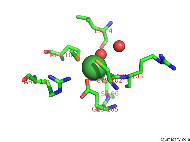
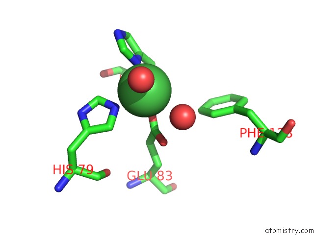
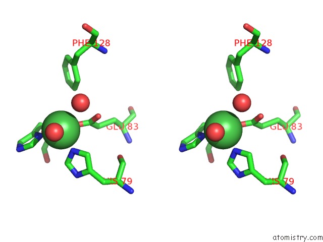
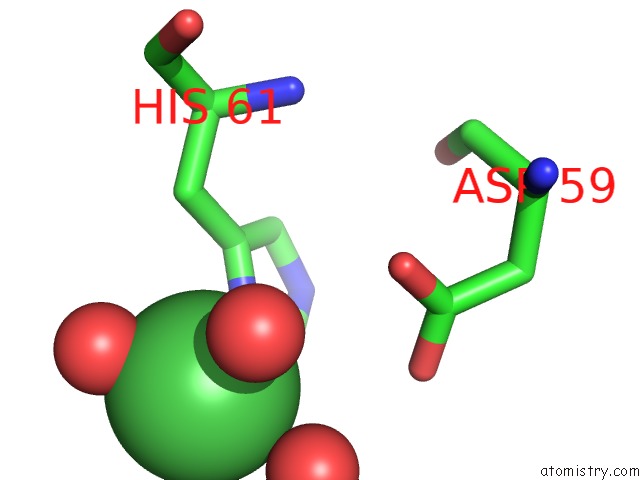
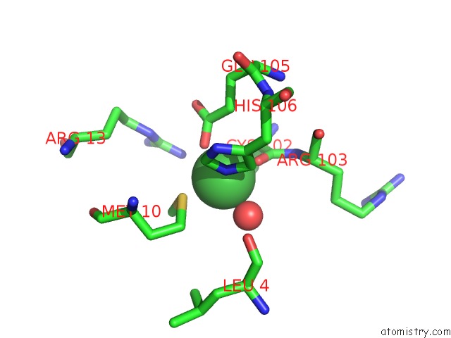
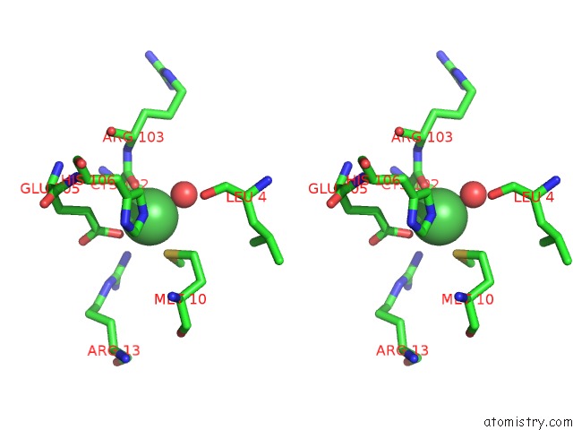
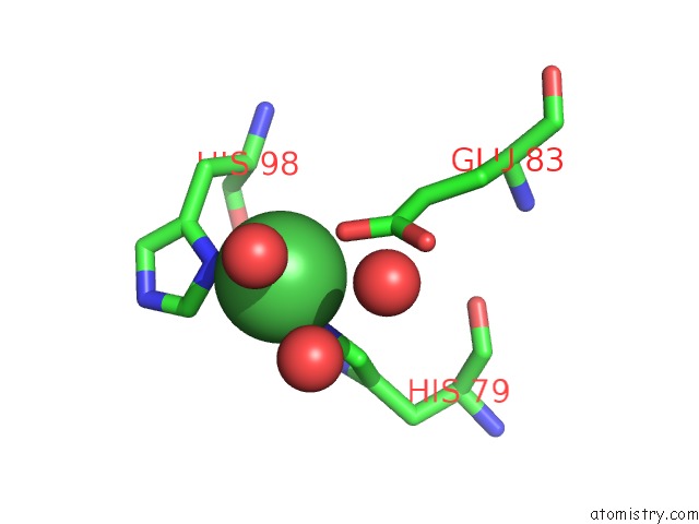
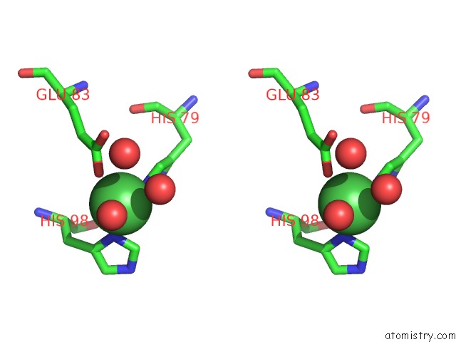
Nickel binding site 2 out of 12 in 2isz
Go back to
Nickel binding site 2 out
of 12 in the Crystal Structure of A Two-Domain Ider-Dna Complex Crystal Form I
Mono view
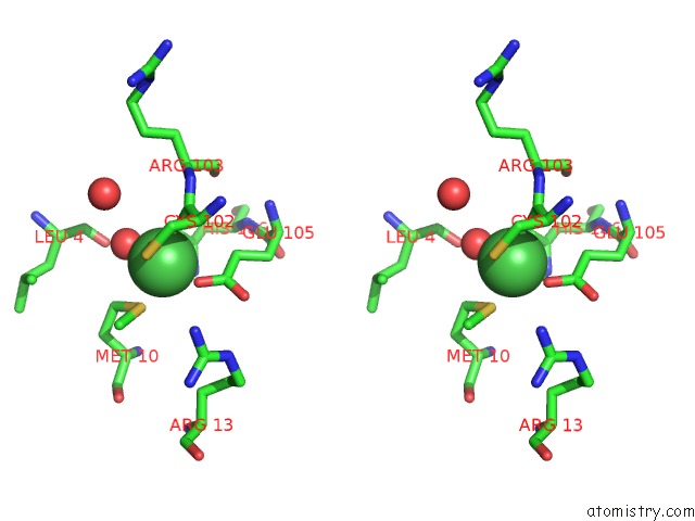
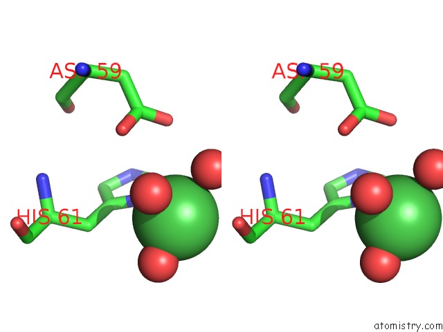
Stereo pair view
Mono view
Stereo pair view
A full contact list of Nickel with other atoms in the Ni binding
site number 2 of Crystal Structure of A Two-Domain Ider-Dna Complex Crystal Form I within 5.0Å range:
|
Nickel binding site 3 out of 12 in 2isz
Go back to
Nickel binding site 3 out
of 12 in the Crystal Structure of A Two-Domain Ider-Dna Complex Crystal Form I
Mono view
Stereo pair view
Mono view
Stereo pair view
A full contact list of Nickel with other atoms in the Ni binding
site number 3 of Crystal Structure of A Two-Domain Ider-Dna Complex Crystal Form I within 5.0Å range:
|
Nickel binding site 4 out of 12 in 2isz
Go back to
Nickel binding site 4 out
of 12 in the Crystal Structure of A Two-Domain Ider-Dna Complex Crystal Form I
Mono view
Stereo pair view
Mono view
Stereo pair view
A full contact list of Nickel with other atoms in the Ni binding
site number 4 of Crystal Structure of A Two-Domain Ider-Dna Complex Crystal Form I within 5.0Å range:
|
Nickel binding site 5 out of 12 in 2isz
Go back to
Nickel binding site 5 out
of 12 in the Crystal Structure of A Two-Domain Ider-Dna Complex Crystal Form I
Mono view
Stereo pair view
Mono view
Stereo pair view
A full contact list of Nickel with other atoms in the Ni binding
site number 5 of Crystal Structure of A Two-Domain Ider-Dna Complex Crystal Form I within 5.0Å range:
|
Nickel binding site 6 out of 12 in 2isz
Go back to
Nickel binding site 6 out
of 12 in the Crystal Structure of A Two-Domain Ider-Dna Complex Crystal Form I
Mono view
Stereo pair view
Mono view
Stereo pair view
A full contact list of Nickel with other atoms in the Ni binding
site number 6 of Crystal Structure of A Two-Domain Ider-Dna Complex Crystal Form I within 5.0Å range:
|
Nickel binding site 7 out of 12 in 2isz
Go back to
Nickel binding site 7 out
of 12 in the Crystal Structure of A Two-Domain Ider-Dna Complex Crystal Form I
Mono view
Stereo pair view
Mono view
Stereo pair view
A full contact list of Nickel with other atoms in the Ni binding
site number 7 of Crystal Structure of A Two-Domain Ider-Dna Complex Crystal Form I within 5.0Å range:
|
Nickel binding site 8 out of 12 in 2isz
Go back to
Nickel binding site 8 out
of 12 in the Crystal Structure of A Two-Domain Ider-Dna Complex Crystal Form I
Mono view
Stereo pair view
Mono view
Stereo pair view
A full contact list of Nickel with other atoms in the Ni binding
site number 8 of Crystal Structure of A Two-Domain Ider-Dna Complex Crystal Form I within 5.0Å range:
|
Nickel binding site 9 out of 12 in 2isz
Go back to
Nickel binding site 9 out
of 12 in the Crystal Structure of A Two-Domain Ider-Dna Complex Crystal Form I
Mono view
Stereo pair view
Mono view
Stereo pair view
A full contact list of Nickel with other atoms in the Ni binding
site number 9 of Crystal Structure of A Two-Domain Ider-Dna Complex Crystal Form I within 5.0Å range:
|
Nickel binding site 10 out of 12 in 2isz
Go back to
Nickel binding site 10 out
of 12 in the Crystal Structure of A Two-Domain Ider-Dna Complex Crystal Form I
Mono view
Stereo pair view
Mono view
Stereo pair view
A full contact list of Nickel with other atoms in the Ni binding
site number 10 of Crystal Structure of A Two-Domain Ider-Dna Complex Crystal Form I within 5.0Å range:
|
Reference:
G.Wisedchaisri,
C.J.Chou,
M.Wu,
C.Roach,
A.E.Rice,
R.K.Holmes,
C.Beeson,
W.G.Hol.
Crystal Structures, Metal Activation, and Dna-Binding Properties of Two-Domain Ider From Mycobacterium Tuberculosis Biochemistry V. 46 436 2007.
ISSN: ISSN 0006-2960
PubMed: 17209554
DOI: 10.1021/BI0609826
Page generated: Wed Oct 9 16:49:04 2024
ISSN: ISSN 0006-2960
PubMed: 17209554
DOI: 10.1021/BI0609826
Last articles
Zn in 9J0NZn in 9J0O
Zn in 9J0P
Zn in 9FJX
Zn in 9EKB
Zn in 9C0F
Zn in 9CAH
Zn in 9CH0
Zn in 9CH3
Zn in 9CH1