Nickel »
PDB 2gw4-2pos »
2ok3 »
Nickel in PDB 2ok3: X-Ray Structure of Human Cyclophilin J at 2.0 Angstrom
Enzymatic activity of X-Ray Structure of Human Cyclophilin J at 2.0 Angstrom
All present enzymatic activity of X-Ray Structure of Human Cyclophilin J at 2.0 Angstrom:
5.2.1.8;
5.2.1.8;
Protein crystallography data
The structure of X-Ray Structure of Human Cyclophilin J at 2.0 Angstrom, PDB code: 2ok3
was solved by
Z.Xia,
with X-Ray Crystallography technique. A brief refinement statistics is given in the table below:
| Resolution Low / High (Å) | 22.11 / 2.00 |
| Space group | P 31 2 1 |
| Cell size a, b, c (Å), α, β, γ (°) | 40.577, 40.577, 170.682, 90.00, 90.00, 120.00 |
| R / Rfree (%) | 20.2 / 25.5 |
Nickel Binding Sites:
The binding sites of Nickel atom in the X-Ray Structure of Human Cyclophilin J at 2.0 Angstrom
(pdb code 2ok3). This binding sites where shown within
5.0 Angstroms radius around Nickel atom.
In total 2 binding sites of Nickel where determined in the X-Ray Structure of Human Cyclophilin J at 2.0 Angstrom, PDB code: 2ok3:
Jump to Nickel binding site number: 1; 2;
In total 2 binding sites of Nickel where determined in the X-Ray Structure of Human Cyclophilin J at 2.0 Angstrom, PDB code: 2ok3:
Jump to Nickel binding site number: 1; 2;
Nickel binding site 1 out of 2 in 2ok3
Go back to
Nickel binding site 1 out
of 2 in the X-Ray Structure of Human Cyclophilin J at 2.0 Angstrom
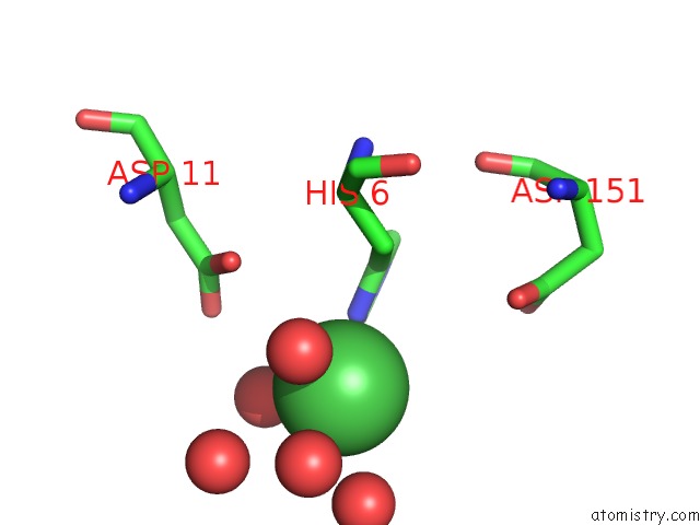
Mono view
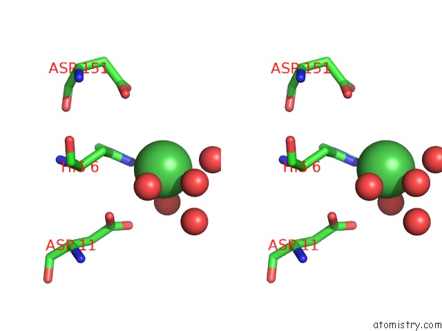
Stereo pair view
Mono view
Stereo pair view
A full contact list of Nickel with other atoms in the Ni binding
site number 1 of X-Ray Structure of Human Cyclophilin J at 2.0 Angstrom within 5.0Å range:
|
Nickel binding site 2 out of 2 in 2ok3
Go back to
Nickel binding site 2 out
of 2 in the X-Ray Structure of Human Cyclophilin J at 2.0 Angstrom
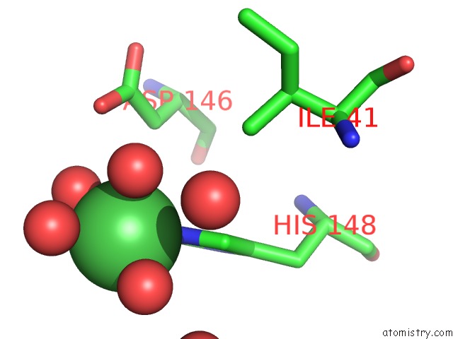
Mono view
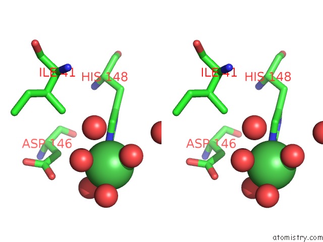
Stereo pair view
Mono view
Stereo pair view
A full contact list of Nickel with other atoms in the Ni binding
site number 2 of X-Ray Structure of Human Cyclophilin J at 2.0 Angstrom within 5.0Å range:
|
Reference:
J.Chen,
S.Chen,
L.Huang,
X.Zhao,
J.Tan,
C.Huang,
H.Saiyin,
M.Zhang,
X.Zeng,
J.Xi,
B.Wan,
Y.Zhao,
Z.Xia,
H.Jiang,
Q.Yi,
J.O.Liu,
L.Yu.
Targeting Cyclophilin J, A Novel Peptidyl-Prolyl Isomerase, Can Induce Cellular G1/S Arrest and Repress the Growth of Hepatocellular Carcinoma To Be Published.
Page generated: Wed Oct 9 16:52:56 2024
Last articles
Al in 4ANJAl in 3WGV
Al in 4A36
Al in 3ZS9
Al in 3WGU
Al in 3SYN
Al in 3W6P
Al in 3T34
Al in 3UKD
Al in 3RFU