Nickel »
PDB 2pr0-2w3u »
2v24 »
Nickel in PDB 2v24: Structure of the Human Spry Domain-Containing Socs Box Protein Ssb-4
Protein crystallography data
The structure of Structure of the Human Spry Domain-Containing Socs Box Protein Ssb-4, PDB code: 2v24
was solved by
J.Uppenberg,
A.Bullock,
T.Keates,
P.Savitsky,
A.C.W.Pike,
E.Ugochukwu,
G.Bunkoczi,
F.Von Delft,
J.Weigelt,
C.H.Arrowsmith,
A.Edwards,
M.Sundstrom,
S.Knapp,
with X-Ray Crystallography technique. A brief refinement statistics is given in the table below:
| Resolution Low / High (Å) | 50.00 / 2.20 |
| Space group | P 32 2 1 |
| Cell size a, b, c (Å), α, β, γ (°) | 104.220, 104.220, 40.780, 90.00, 90.00, 120.00 |
| R / Rfree (%) | 20.9 / 26 |
Nickel Binding Sites:
The binding sites of Nickel atom in the Structure of the Human Spry Domain-Containing Socs Box Protein Ssb-4
(pdb code 2v24). This binding sites where shown within
5.0 Angstroms radius around Nickel atom.
In total only one binding site of Nickel was determined in the Structure of the Human Spry Domain-Containing Socs Box Protein Ssb-4, PDB code: 2v24:
In total only one binding site of Nickel was determined in the Structure of the Human Spry Domain-Containing Socs Box Protein Ssb-4, PDB code: 2v24:
Nickel binding site 1 out of 1 in 2v24
Go back to
Nickel binding site 1 out
of 1 in the Structure of the Human Spry Domain-Containing Socs Box Protein Ssb-4
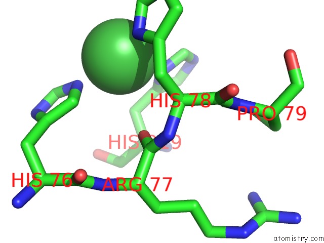
Mono view
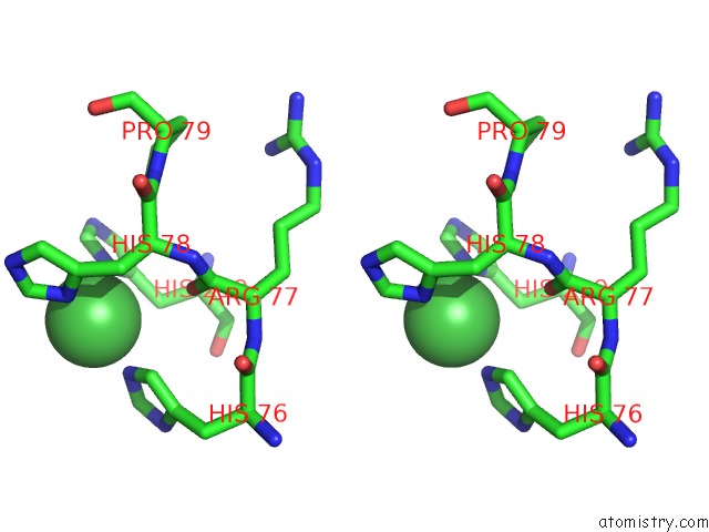
Stereo pair view
Mono view
Stereo pair view
A full contact list of Nickel with other atoms in the Ni binding
site number 1 of Structure of the Human Spry Domain-Containing Socs Box Protein Ssb-4 within 5.0Å range:
|
Reference:
P.Filippakopoulos,
A.Low,
T.D.Sharpe,
J.Uppenberg,
S.Yao,
Z.Kuang,
P.Savitsky,
R.S.Lewis,
S.E.Nicholson,
R.S.Norton,
A.Bullock.
Structural Basis For Par-4 Recognition By the Spry Domain- and Socs Box-Containing Proteins SPSB1, SPSB2, and SPSB4. J.Mol.Biol. V. 401 389 2010.
ISSN: ISSN 0022-2836
PubMed: 20561531
DOI: 10.1016/J.JMB.2010.06.017
Page generated: Wed Oct 9 16:59:09 2024
ISSN: ISSN 0022-2836
PubMed: 20561531
DOI: 10.1016/J.JMB.2010.06.017
Last articles
Zn in 9MJ5Zn in 9HNW
Zn in 9G0L
Zn in 9FNE
Zn in 9DZN
Zn in 9E0I
Zn in 9D32
Zn in 9DAK
Zn in 8ZXC
Zn in 8ZUF