Nickel »
PDB 2pr0-2w3u »
2w2i »
Nickel in PDB 2w2i: Crystal Structure of the Human 2-Oxoglutarate Oxygenase LOC390245
Protein crystallography data
The structure of Crystal Structure of the Human 2-Oxoglutarate Oxygenase LOC390245, PDB code: 2w2i
was solved by
W.W.Yue,
S.Ng,
N.Shafqat,
E.Ugochukwu,
M.Mcdonough,
A.C.W.Pike,
P.Filippakopoulos,
F.Von Delft,
C.Arrowsmith,
J.Weigelt,
A.Edwards,
C.Bountra,
C.Schofield,
U.Oppermann,
with X-Ray Crystallography technique. A brief refinement statistics is given in the table below:
| Resolution Low / High (Å) | 111.80 / 2.1 |
| Space group | I 2 2 2 |
| Cell size a, b, c (Å), α, β, γ (°) | 90.904, 111.009, 224.199, 90.00, 90.00, 90.00 |
| R / Rfree (%) | 21 / 23.6 |
Nickel Binding Sites:
The binding sites of Nickel atom in the Crystal Structure of the Human 2-Oxoglutarate Oxygenase LOC390245
(pdb code 2w2i). This binding sites where shown within
5.0 Angstroms radius around Nickel atom.
In total 3 binding sites of Nickel where determined in the Crystal Structure of the Human 2-Oxoglutarate Oxygenase LOC390245, PDB code: 2w2i:
Jump to Nickel binding site number: 1; 2; 3;
In total 3 binding sites of Nickel where determined in the Crystal Structure of the Human 2-Oxoglutarate Oxygenase LOC390245, PDB code: 2w2i:
Jump to Nickel binding site number: 1; 2; 3;
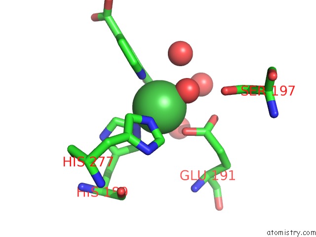
Nickel binding site 1 out of 3 in 2w2i
Go back to
Nickel binding site 1 out
of 3 in the Crystal Structure of the Human 2-Oxoglutarate Oxygenase LOC390245

Mono view

Stereo pair view
Mono view
Stereo pair view
A full contact list of Nickel with other atoms in the Ni binding
site number 1 of Crystal Structure of the Human 2-Oxoglutarate Oxygenase LOC390245 within 5.0Å range:
|
Nickel binding site 2 out of 3 in 2w2i
Go back to
Nickel binding site 2 out
of 3 in the Crystal Structure of the Human 2-Oxoglutarate Oxygenase LOC390245
Mono view

Stereo pair view
Mono view
Stereo pair view
A full contact list of Nickel with other atoms in the Ni binding
site number 2 of Crystal Structure of the Human 2-Oxoglutarate Oxygenase LOC390245 within 5.0Å range:
|
Nickel binding site 3 out of 3 in 2w2i
Go back to
Nickel binding site 3 out
of 3 in the Crystal Structure of the Human 2-Oxoglutarate Oxygenase LOC390245

Mono view

Stereo pair view
Mono view
Stereo pair view
A full contact list of Nickel with other atoms in the Ni binding
site number 3 of Crystal Structure of the Human 2-Oxoglutarate Oxygenase LOC390245 within 5.0Å range:
|
Reference:
L.Hillringhaus,
W.W.Yue,
N.R.Rose,
S.S.Ng,
C.Gileadi,
C.Loenarz,
S.H.Bello,
J.E.Bray,
C.J.Schofield,
U.Oppermann.
Structural and Evolutionary Basis For the Dual Substrate Selectivity of Human KDM4 Histone Demethylase Family. J.Biol.Chem. V. 286 41616 2011.
ISSN: ISSN 0021-9258
PubMed: 21914792
DOI: 10.1074/JBC.M111.283689
Page generated: Wed Oct 9 17:00:24 2024
ISSN: ISSN 0021-9258
PubMed: 21914792
DOI: 10.1074/JBC.M111.283689
Last articles
Zn in 9MJ5Zn in 9HNW
Zn in 9G0L
Zn in 9FNE
Zn in 9DZN
Zn in 9E0I
Zn in 9D32
Zn in 9DAK
Zn in 8ZXC
Zn in 8ZUF