Nickel »
PDB 2w94-2zpl »
2yjl »
Nickel in PDB 2yjl: Structural Characterization of A Secretin Pilot Protein From the Type III Secretion System (T3SS) of Pseudomonas Aeruginosa
Protein crystallography data
The structure of Structural Characterization of A Secretin Pilot Protein From the Type III Secretion System (T3SS) of Pseudomonas Aeruginosa, PDB code: 2yjl
was solved by
T.Izore,
C.Perdu,
V.Job,
I.Atree,
E.Faudry,
A.Dessen,
with X-Ray Crystallography technique. A brief refinement statistics is given in the table below:
| Resolution Low / High (Å) | 35.54 / 1.81 |
| Space group | C 1 2 1 |
| Cell size a, b, c (Å), α, β, γ (°) | 124.155, 48.358, 71.602, 90.00, 96.92, 90.00 |
| R / Rfree (%) | 21.691 / 26.224 |
Nickel Binding Sites:
The binding sites of Nickel atom in the Structural Characterization of A Secretin Pilot Protein From the Type III Secretion System (T3SS) of Pseudomonas Aeruginosa
(pdb code 2yjl). This binding sites where shown within
5.0 Angstroms radius around Nickel atom.
In total only one binding site of Nickel was determined in the Structural Characterization of A Secretin Pilot Protein From the Type III Secretion System (T3SS) of Pseudomonas Aeruginosa, PDB code: 2yjl:
In total only one binding site of Nickel was determined in the Structural Characterization of A Secretin Pilot Protein From the Type III Secretion System (T3SS) of Pseudomonas Aeruginosa, PDB code: 2yjl:
Nickel binding site 1 out of 1 in 2yjl
Go back to
Nickel binding site 1 out
of 1 in the Structural Characterization of A Secretin Pilot Protein From the Type III Secretion System (T3SS) of Pseudomonas Aeruginosa
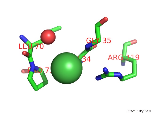
Mono view
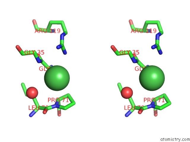
Stereo pair view
Mono view
Stereo pair view
A full contact list of Nickel with other atoms in the Ni binding
site number 1 of Structural Characterization of A Secretin Pilot Protein From the Type III Secretion System (T3SS) of Pseudomonas Aeruginosa within 5.0Å range:
|
Reference:
T.Izore,
C.Perdu,
V.Job,
I.Atree,
E.Faudry,
A.Dessen.
Structural Characterization and Membrane Localization of Exsb From the Type III Secretion System (T3SS) of Pseudomonas Aeruginosa J.Mol.Biol. V. 413 236 2011.
ISSN: ISSN 0022-2836
PubMed: 21839744
DOI: 10.1016/J.JMB.2011.07.043
Page generated: Wed Oct 9 17:07:47 2024
ISSN: ISSN 0022-2836
PubMed: 21839744
DOI: 10.1016/J.JMB.2011.07.043
Last articles
Zn in 9J0NZn in 9J0O
Zn in 9J0P
Zn in 9FJX
Zn in 9EKB
Zn in 9C0F
Zn in 9CAH
Zn in 9CH0
Zn in 9CH3
Zn in 9CH1