Nickel »
PDB 3dse-3hy3 »
3fpu »
Nickel in PDB 3fpu: The Crystallographic Structure of the Complex Between Evasin-1 and CCL3
Protein crystallography data
The structure of The Crystallographic Structure of the Complex Between Evasin-1 and CCL3, PDB code: 3fpu
was solved by
J.P.Shaw,
J.M.Dias,
with X-Ray Crystallography technique. A brief refinement statistics is given in the table below:
| Resolution Low / High (Å) | 50.00 / 1.76 |
| Space group | P 43 3 2 |
| Cell size a, b, c (Å), α, β, γ (°) | 104.384, 104.384, 104.384, 90.00, 90.00, 90.00 |
| R / Rfree (%) | 23.1 / 28.5 |
Nickel Binding Sites:
The binding sites of Nickel atom in the The Crystallographic Structure of the Complex Between Evasin-1 and CCL3
(pdb code 3fpu). This binding sites where shown within
5.0 Angstroms radius around Nickel atom.
In total 2 binding sites of Nickel where determined in the The Crystallographic Structure of the Complex Between Evasin-1 and CCL3, PDB code: 3fpu:
Jump to Nickel binding site number: 1; 2;
In total 2 binding sites of Nickel where determined in the The Crystallographic Structure of the Complex Between Evasin-1 and CCL3, PDB code: 3fpu:
Jump to Nickel binding site number: 1; 2;
Nickel binding site 1 out of 2 in 3fpu
Go back to
Nickel binding site 1 out
of 2 in the The Crystallographic Structure of the Complex Between Evasin-1 and CCL3
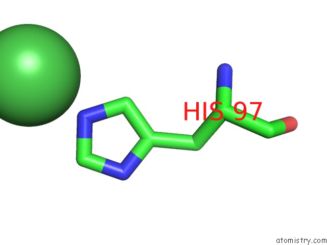
Mono view
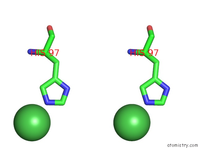
Stereo pair view
Mono view
Stereo pair view
A full contact list of Nickel with other atoms in the Ni binding
site number 1 of The Crystallographic Structure of the Complex Between Evasin-1 and CCL3 within 5.0Å range:
|
Nickel binding site 2 out of 2 in 3fpu
Go back to
Nickel binding site 2 out
of 2 in the The Crystallographic Structure of the Complex Between Evasin-1 and CCL3
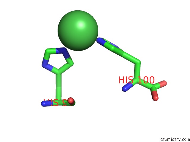
Mono view
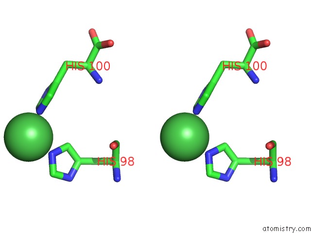
Stereo pair view
Mono view
Stereo pair view
A full contact list of Nickel with other atoms in the Ni binding
site number 2 of The Crystallographic Structure of the Complex Between Evasin-1 and CCL3 within 5.0Å range:
|
Reference:
J.M.Dias,
C.Losberger,
M.Deruaz,
C.A.Power,
A.E.I.Proudfoot,
J.P.Shaw.
Structural Basis of Chemokine Sequestration By A Tick Chemokine Binding Protein: the Crystal Structure of the Complex Between Evasin-1 and CCL3 Plos One V. 4 2009.
ISSN: ESSN 1932-6203
PubMed: 20041127
DOI: 10.1371/JOURNAL.PONE.0008514
Page generated: Wed Oct 9 17:17:20 2024
ISSN: ESSN 1932-6203
PubMed: 20041127
DOI: 10.1371/JOURNAL.PONE.0008514
Last articles
Zn in 9MJ5Zn in 9HNW
Zn in 9G0L
Zn in 9FNE
Zn in 9DZN
Zn in 9E0I
Zn in 9D32
Zn in 9DAK
Zn in 8ZXC
Zn in 8ZUF