Nickel »
PDB 3dse-3hy3 »
3gw7 »
Nickel in PDB 3gw7: Crystal Structure of A Metal-Dependent Phosphohydrolase with Conserved Hd Domain (Yedj) From Escherichia Coli in Complex with Nickel Ions. Northeast Structural Genomics Consortium Target ER63
Protein crystallography data
The structure of Crystal Structure of A Metal-Dependent Phosphohydrolase with Conserved Hd Domain (Yedj) From Escherichia Coli in Complex with Nickel Ions. Northeast Structural Genomics Consortium Target ER63, PDB code: 3gw7
was solved by
F.Forouhar,
M.Abashidze,
J.Seetharaman,
J.Janjua,
R.Xiao,
K.Cunningham,
L.Ma,
L.Zhao,
J.K.Everett,
R.Nair,
T.B.Acton,
B.Rost,
G.T.Montelione,
J.F.Hunt,
L.Tong,
Northeast Structural Genomics Consortium (Nesg),
with X-Ray Crystallography technique. A brief refinement statistics is given in the table below:
| Resolution Low / High (Å) | 20.00 / 3.30 |
| Space group | P 63 |
| Cell size a, b, c (Å), α, β, γ (°) | 99.408, 99.408, 130.629, 90.00, 90.00, 120.00 |
| R / Rfree (%) | 26 / 29.4 |
Nickel Binding Sites:
The binding sites of Nickel atom in the Crystal Structure of A Metal-Dependent Phosphohydrolase with Conserved Hd Domain (Yedj) From Escherichia Coli in Complex with Nickel Ions. Northeast Structural Genomics Consortium Target ER63
(pdb code 3gw7). This binding sites where shown within
5.0 Angstroms radius around Nickel atom.
In total 8 binding sites of Nickel where determined in the Crystal Structure of A Metal-Dependent Phosphohydrolase with Conserved Hd Domain (Yedj) From Escherichia Coli in Complex with Nickel Ions. Northeast Structural Genomics Consortium Target ER63, PDB code: 3gw7:
Jump to Nickel binding site number: 1; 2; 3; 4; 5; 6; 7; 8;
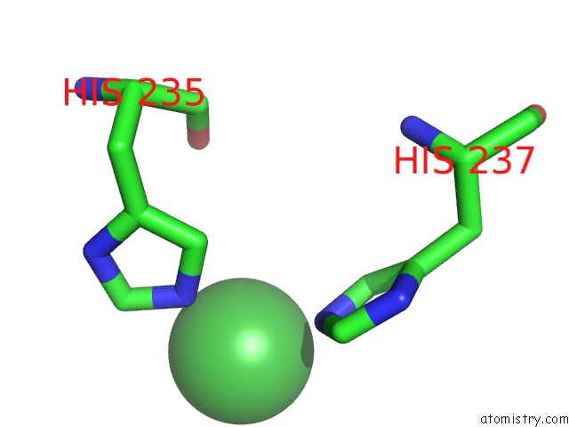
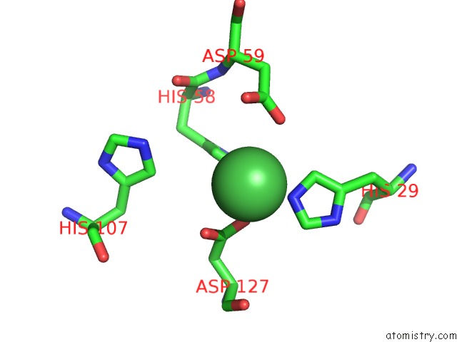
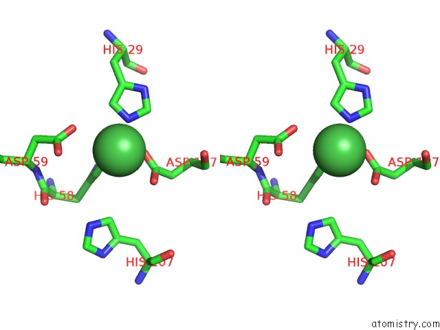
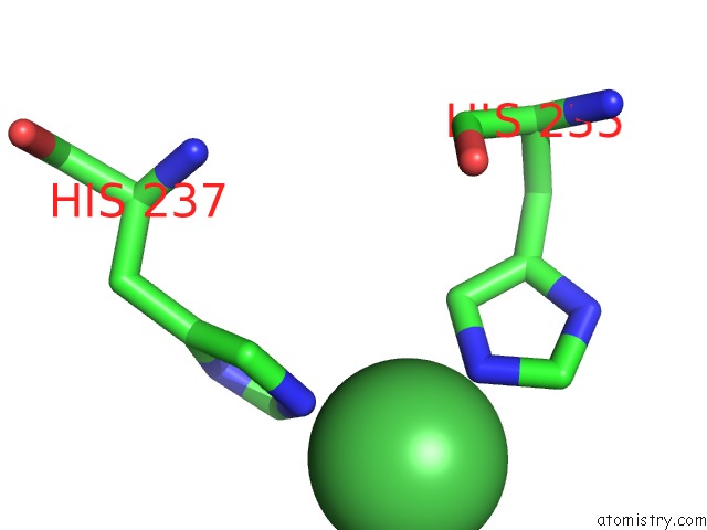
Nickel binding site 1 out of 8 in 3gw7
Go back to
Nickel binding site 1 out
of 8 in the Crystal Structure of A Metal-Dependent Phosphohydrolase with Conserved Hd Domain (Yedj) From Escherichia Coli in Complex with Nickel Ions. Northeast Structural Genomics Consortium Target ER63
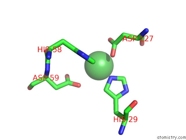
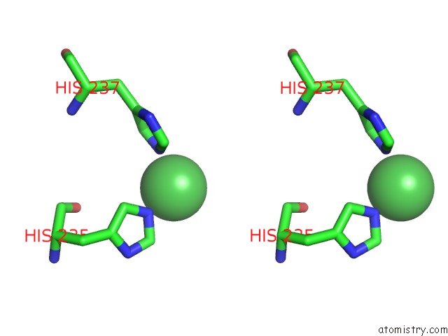
Mono view
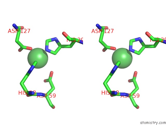
Stereo pair view
Mono view
Stereo pair view
A full contact list of Nickel with other atoms in the Ni binding
site number 1 of Crystal Structure of A Metal-Dependent Phosphohydrolase with Conserved Hd Domain (Yedj) From Escherichia Coli in Complex with Nickel Ions. Northeast Structural Genomics Consortium Target ER63 within 5.0Å range:
|
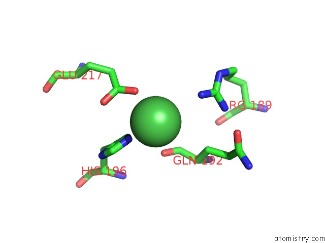
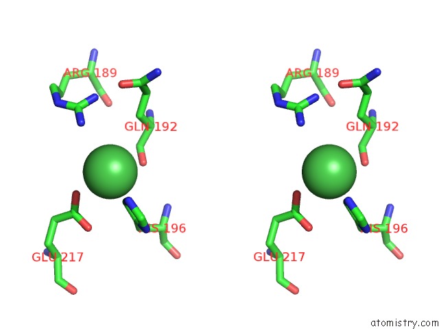
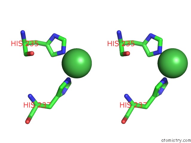
Nickel binding site 2 out of 8 in 3gw7
Go back to
Nickel binding site 2 out
of 8 in the Crystal Structure of A Metal-Dependent Phosphohydrolase with Conserved Hd Domain (Yedj) From Escherichia Coli in Complex with Nickel Ions. Northeast Structural Genomics Consortium Target ER63
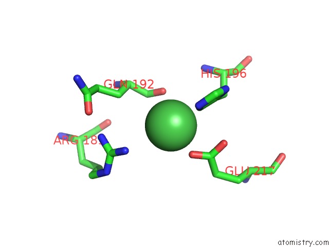
Mono view
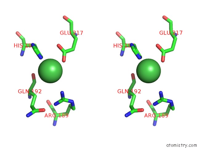
Stereo pair view
Mono view
Stereo pair view
A full contact list of Nickel with other atoms in the Ni binding
site number 2 of Crystal Structure of A Metal-Dependent Phosphohydrolase with Conserved Hd Domain (Yedj) From Escherichia Coli in Complex with Nickel Ions. Northeast Structural Genomics Consortium Target ER63 within 5.0Å range:
|
Nickel binding site 3 out of 8 in 3gw7
Go back to
Nickel binding site 3 out
of 8 in the Crystal Structure of A Metal-Dependent Phosphohydrolase with Conserved Hd Domain (Yedj) From Escherichia Coli in Complex with Nickel Ions. Northeast Structural Genomics Consortium Target ER63
Mono view
Stereo pair view
Mono view
Stereo pair view
A full contact list of Nickel with other atoms in the Ni binding
site number 3 of Crystal Structure of A Metal-Dependent Phosphohydrolase with Conserved Hd Domain (Yedj) From Escherichia Coli in Complex with Nickel Ions. Northeast Structural Genomics Consortium Target ER63 within 5.0Å range:
|
Nickel binding site 4 out of 8 in 3gw7
Go back to
Nickel binding site 4 out
of 8 in the Crystal Structure of A Metal-Dependent Phosphohydrolase with Conserved Hd Domain (Yedj) From Escherichia Coli in Complex with Nickel Ions. Northeast Structural Genomics Consortium Target ER63
Mono view
Stereo pair view
Mono view
Stereo pair view
A full contact list of Nickel with other atoms in the Ni binding
site number 4 of Crystal Structure of A Metal-Dependent Phosphohydrolase with Conserved Hd Domain (Yedj) From Escherichia Coli in Complex with Nickel Ions. Northeast Structural Genomics Consortium Target ER63 within 5.0Å range:
|
Nickel binding site 5 out of 8 in 3gw7
Go back to
Nickel binding site 5 out
of 8 in the Crystal Structure of A Metal-Dependent Phosphohydrolase with Conserved Hd Domain (Yedj) From Escherichia Coli in Complex with Nickel Ions. Northeast Structural Genomics Consortium Target ER63
Mono view
Stereo pair view
Mono view
Stereo pair view
A full contact list of Nickel with other atoms in the Ni binding
site number 5 of Crystal Structure of A Metal-Dependent Phosphohydrolase with Conserved Hd Domain (Yedj) From Escherichia Coli in Complex with Nickel Ions. Northeast Structural Genomics Consortium Target ER63 within 5.0Å range:
|
Nickel binding site 6 out of 8 in 3gw7
Go back to
Nickel binding site 6 out
of 8 in the Crystal Structure of A Metal-Dependent Phosphohydrolase with Conserved Hd Domain (Yedj) From Escherichia Coli in Complex with Nickel Ions. Northeast Structural Genomics Consortium Target ER63
Mono view
Stereo pair view
Mono view
Stereo pair view
A full contact list of Nickel with other atoms in the Ni binding
site number 6 of Crystal Structure of A Metal-Dependent Phosphohydrolase with Conserved Hd Domain (Yedj) From Escherichia Coli in Complex with Nickel Ions. Northeast Structural Genomics Consortium Target ER63 within 5.0Å range:
|
Nickel binding site 7 out of 8 in 3gw7
Go back to
Nickel binding site 7 out
of 8 in the Crystal Structure of A Metal-Dependent Phosphohydrolase with Conserved Hd Domain (Yedj) From Escherichia Coli in Complex with Nickel Ions. Northeast Structural Genomics Consortium Target ER63
Mono view
Stereo pair view
Mono view
Stereo pair view
A full contact list of Nickel with other atoms in the Ni binding
site number 7 of Crystal Structure of A Metal-Dependent Phosphohydrolase with Conserved Hd Domain (Yedj) From Escherichia Coli in Complex with Nickel Ions. Northeast Structural Genomics Consortium Target ER63 within 5.0Å range:
|
Nickel binding site 8 out of 8 in 3gw7
Go back to
Nickel binding site 8 out
of 8 in the Crystal Structure of A Metal-Dependent Phosphohydrolase with Conserved Hd Domain (Yedj) From Escherichia Coli in Complex with Nickel Ions. Northeast Structural Genomics Consortium Target ER63
Mono view
Stereo pair view
Mono view
Stereo pair view
A full contact list of Nickel with other atoms in the Ni binding
site number 8 of Crystal Structure of A Metal-Dependent Phosphohydrolase with Conserved Hd Domain (Yedj) From Escherichia Coli in Complex with Nickel Ions. Northeast Structural Genomics Consortium Target ER63 within 5.0Å range:
|
Reference:
F.Forouhar,
M.Abashidze,
J.Seetharaman,
J.Janjua,
R.Xiao,
K.Cunningham,
L.Ma,
L.Zhao,
J.K.Everett,
R.Nair,
T.B.Acton,
B.Rost,
G.T.Montelione,
J.F.Hunt,
L.Tong.
Crystal Structure of A Metal-Dependent Phosphohydrolase with Conserved Hd Domain (Yedj) From Escherichia Coli in Complex with Nickel Ions. Northeast Structural Genomics Consortium Target ER63 To Be Published.
Page generated: Wed Oct 9 17:19:16 2024
Last articles
Zn in 9J0NZn in 9J0O
Zn in 9J0P
Zn in 9FJX
Zn in 9EKB
Zn in 9C0F
Zn in 9CAH
Zn in 9CH0
Zn in 9CH3
Zn in 9CH1