Nickel »
PDB 3dse-3hy3 »
3hg9 »
Nickel in PDB 3hg9: Crystal Structure of Putative Pilm Protein From Pseudomonas Aeruginosa 2192
Protein crystallography data
The structure of Crystal Structure of Putative Pilm Protein From Pseudomonas Aeruginosa 2192, PDB code: 3hg9
was solved by
V.N.Malashkevich,
R.Toro,
J.M.Sauder,
S.K.Burley,
S.C.Almo,
New York Sgxresearch Center For Structural Genomics (Nysgxrc),
with X-Ray Crystallography technique. A brief refinement statistics is given in the table below:
| Resolution Low / High (Å) | 50.00 / 2.40 |
| Space group | P 62 |
| Cell size a, b, c (Å), α, β, γ (°) | 64.516, 64.516, 143.134, 90.00, 90.00, 120.00 |
| R / Rfree (%) | 23.5 / 26.7 |
Nickel Binding Sites:
The binding sites of Nickel atom in the Crystal Structure of Putative Pilm Protein From Pseudomonas Aeruginosa 2192
(pdb code 3hg9). This binding sites where shown within
5.0 Angstroms radius around Nickel atom.
In total 2 binding sites of Nickel where determined in the Crystal Structure of Putative Pilm Protein From Pseudomonas Aeruginosa 2192, PDB code: 3hg9:
Jump to Nickel binding site number: 1; 2;
In total 2 binding sites of Nickel where determined in the Crystal Structure of Putative Pilm Protein From Pseudomonas Aeruginosa 2192, PDB code: 3hg9:
Jump to Nickel binding site number: 1; 2;
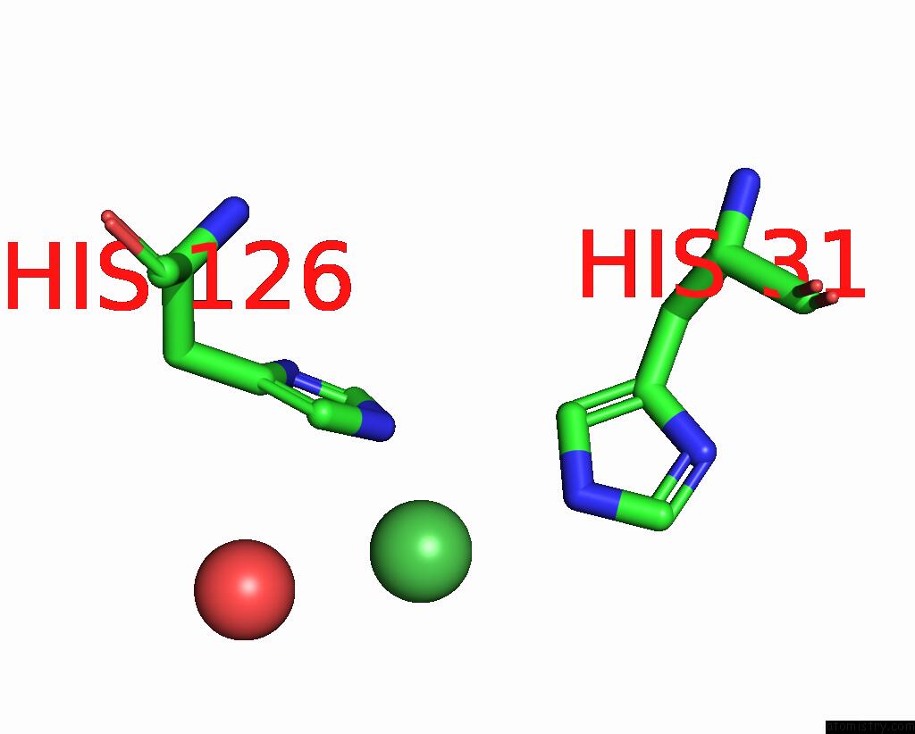
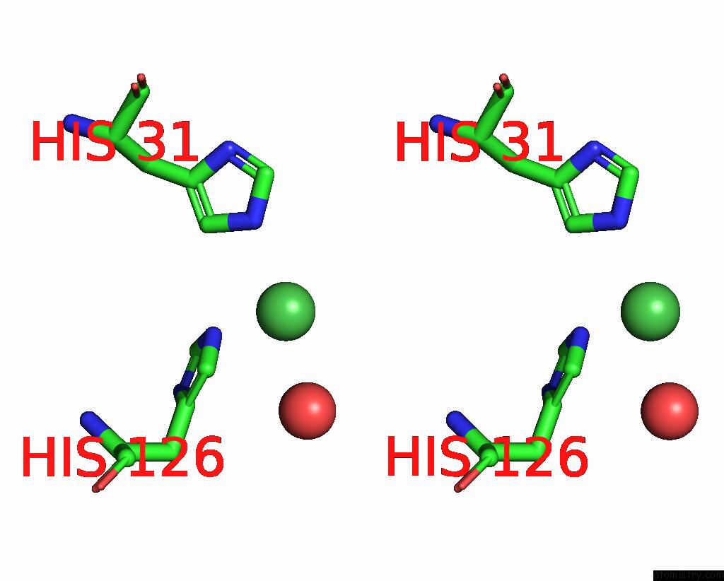
Nickel binding site 1 out of 2 in 3hg9
Go back to
Nickel binding site 1 out
of 2 in the Crystal Structure of Putative Pilm Protein From Pseudomonas Aeruginosa 2192

Mono view

Stereo pair view
Mono view
Stereo pair view
A full contact list of Nickel with other atoms in the Ni binding
site number 1 of Crystal Structure of Putative Pilm Protein From Pseudomonas Aeruginosa 2192 within 5.0Å range:
|
Nickel binding site 2 out of 2 in 3hg9
Go back to
Nickel binding site 2 out
of 2 in the Crystal Structure of Putative Pilm Protein From Pseudomonas Aeruginosa 2192
Mono view
Stereo pair view
Mono view
Stereo pair view
A full contact list of Nickel with other atoms in the Ni binding
site number 2 of Crystal Structure of Putative Pilm Protein From Pseudomonas Aeruginosa 2192 within 5.0Å range:
|
Reference:
V.N.Malashkevich,
R.Toro,
J.M.Sauder,
S.K.Burley,
S.C.Almo.
Crystal Structure of Putative Pilm Protein From Pseudomonas Aeruginosa 2192 To Be Published.
Page generated: Wed Oct 9 17:19:54 2024
Last articles
Al in 9BGHAl in 8XAK
Al in 8WA5
Al in 8UQO
Al in 8VMF
Al in 8SHT
Al in 8UK9
Al in 8SHQ
Al in 8UQN
Al in 8TSI