Nickel »
PDB 3dse-3hy3 »
3ht1 »
Nickel in PDB 3ht1: 1.2A Structure of the Polyketide Cyclase Remf From Streptomyces Resistomycificus
Protein crystallography data
The structure of 1.2A Structure of the Polyketide Cyclase Remf From Streptomyces Resistomycificus, PDB code: 3ht1
was solved by
L.Silvennoinen,
T.Sandalova,
G.Schneider,
with X-Ray Crystallography technique. A brief refinement statistics is given in the table below:
| Resolution Low / High (Å) | 40.00 / 1.20 |
| Space group | P 2 2 21 |
| Cell size a, b, c (Å), α, β, γ (°) | 34.471, 47.907, 96.946, 90.00, 90.00, 90.00 |
| R / Rfree (%) | 12.9 / 17 |
Nickel Binding Sites:
The binding sites of Nickel atom in the 1.2A Structure of the Polyketide Cyclase Remf From Streptomyces Resistomycificus
(pdb code 3ht1). This binding sites where shown within
5.0 Angstroms radius around Nickel atom.
In total only one binding site of Nickel was determined in the 1.2A Structure of the Polyketide Cyclase Remf From Streptomyces Resistomycificus, PDB code: 3ht1:
In total only one binding site of Nickel was determined in the 1.2A Structure of the Polyketide Cyclase Remf From Streptomyces Resistomycificus, PDB code: 3ht1:
Nickel binding site 1 out of 1 in 3ht1
Go back to
Nickel binding site 1 out
of 1 in the 1.2A Structure of the Polyketide Cyclase Remf From Streptomyces Resistomycificus

Mono view
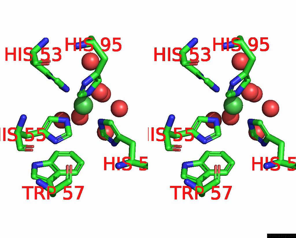
Stereo pair view
Mono view
Stereo pair view
A full contact list of Nickel with other atoms in the Ni binding
site number 1 of 1.2A Structure of the Polyketide Cyclase Remf From Streptomyces Resistomycificus within 5.0Å range:
|
Reference:
L.Silvennoinen,
T.Sandalova,
G.Schneider.
The Polyketide Cyclase Remf From Streptomyces Resistomycificus Contains An Unusual Octahedral Zinc Binding Site Febs Lett. V. 583 2917 2009.
ISSN: ISSN 0014-5793
PubMed: 19665022
DOI: 10.1016/J.FEBSLET.2009.07.061
Page generated: Wed Oct 9 17:20:06 2024
ISSN: ISSN 0014-5793
PubMed: 19665022
DOI: 10.1016/J.FEBSLET.2009.07.061
Last articles
Zn in 9MJ5Zn in 9HNW
Zn in 9G0L
Zn in 9FNE
Zn in 9DZN
Zn in 9E0I
Zn in 9D32
Zn in 9DAK
Zn in 8ZXC
Zn in 8ZUF