Nickel »
PDB 3nbk-3qsf »
3ny0 »
Nickel in PDB 3ny0: Crystal Structure of Uree From Helicobacter Pylori (NI2+ Bound Form)
Protein crystallography data
The structure of Crystal Structure of Uree From Helicobacter Pylori (NI2+ Bound Form), PDB code: 3ny0
was solved by
R.Shi,
C.Munger,
A.Assinas,
A.Matte,
M.Cygler,
Montreal-Kingston Bacterialstructural Genomics Initiative (Bsgi),
with X-Ray Crystallography technique. A brief refinement statistics is given in the table below:
| Resolution Low / High (Å) | 46.63 / 3.09 |
| Space group | P 41 21 2 |
| Cell size a, b, c (Å), α, β, γ (°) | 91.057, 91.057, 202.823, 90.00, 90.00, 90.00 |
| R / Rfree (%) | 24.5 / 29.9 |
Nickel Binding Sites:
The binding sites of Nickel atom in the Crystal Structure of Uree From Helicobacter Pylori (NI2+ Bound Form)
(pdb code 3ny0). This binding sites where shown within
5.0 Angstroms radius around Nickel atom.
In total only one binding site of Nickel was determined in the Crystal Structure of Uree From Helicobacter Pylori (NI2+ Bound Form), PDB code: 3ny0:
In total only one binding site of Nickel was determined in the Crystal Structure of Uree From Helicobacter Pylori (NI2+ Bound Form), PDB code: 3ny0:
Nickel binding site 1 out of 1 in 3ny0
Go back to
Nickel binding site 1 out
of 1 in the Crystal Structure of Uree From Helicobacter Pylori (NI2+ Bound Form)
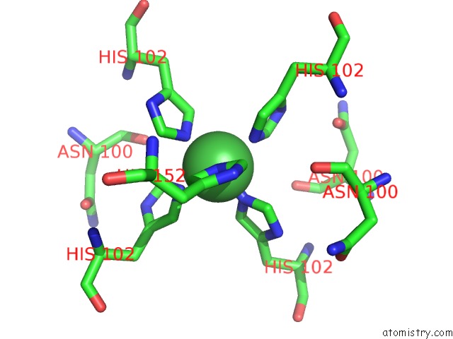
Mono view
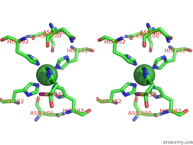
Stereo pair view
Mono view
Stereo pair view
A full contact list of Nickel with other atoms in the Ni binding
site number 1 of Crystal Structure of Uree From Helicobacter Pylori (NI2+ Bound Form) within 5.0Å range:
|
Reference:
R.Shi,
C.Munger,
A.Asinas,
S.L.Benoit,
E.Miller,
A.Matte,
R.J.Maier,
M.Cygler.
Crystal Structures of Apo and Metal-Bound Forms of the Uree Protein From Helicobacter Pylori: Role of Multiple Metal Binding Sites Biochemistry V. 49 7080 2010.
ISSN: ISSN 0006-2960
PubMed: 20681615
DOI: 10.1021/BI100372H
Page generated: Wed Oct 9 17:40:18 2024
ISSN: ISSN 0006-2960
PubMed: 20681615
DOI: 10.1021/BI100372H
Last articles
Zn in 9MJ5Zn in 9HNW
Zn in 9G0L
Zn in 9FNE
Zn in 9DZN
Zn in 9E0I
Zn in 9D32
Zn in 9DAK
Zn in 8ZXC
Zn in 8ZUF