Nickel »
PDB 3nbk-3qsf »
3pht »
Nickel in PDB 3pht: Crystal Structure of H74A Mutant of Helicobacter Pylori Nikr
Protein crystallography data
The structure of Crystal Structure of H74A Mutant of Helicobacter Pylori Nikr, PDB code: 3pht
was solved by
E.Pozharski,
S.Evans,
S.Michel,
with X-Ray Crystallography technique. A brief refinement statistics is given in the table below:
| Resolution Low / High (Å) | 61.65 / 2.04 |
| Space group | P 43 21 2 |
| Cell size a, b, c (Å), α, β, γ (°) | 73.174, 73.174, 114.441, 90.00, 90.00, 90.00 |
| R / Rfree (%) | 19.1 / 23 |
Nickel Binding Sites:
The binding sites of Nickel atom in the Crystal Structure of H74A Mutant of Helicobacter Pylori Nikr
(pdb code 3pht). This binding sites where shown within
5.0 Angstroms radius around Nickel atom.
In total 2 binding sites of Nickel where determined in the Crystal Structure of H74A Mutant of Helicobacter Pylori Nikr, PDB code: 3pht:
Jump to Nickel binding site number: 1; 2;
In total 2 binding sites of Nickel where determined in the Crystal Structure of H74A Mutant of Helicobacter Pylori Nikr, PDB code: 3pht:
Jump to Nickel binding site number: 1; 2;
Nickel binding site 1 out of 2 in 3pht
Go back to
Nickel binding site 1 out
of 2 in the Crystal Structure of H74A Mutant of Helicobacter Pylori Nikr
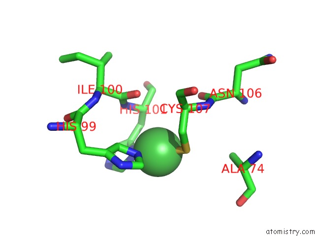
Mono view
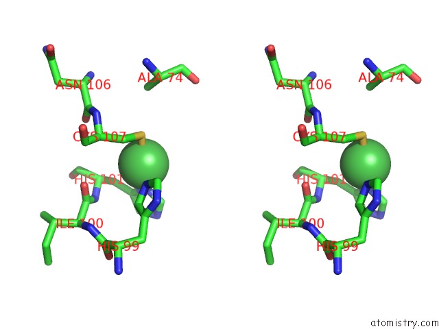
Stereo pair view
Mono view
Stereo pair view
A full contact list of Nickel with other atoms in the Ni binding
site number 1 of Crystal Structure of H74A Mutant of Helicobacter Pylori Nikr within 5.0Å range:
|
Nickel binding site 2 out of 2 in 3pht
Go back to
Nickel binding site 2 out
of 2 in the Crystal Structure of H74A Mutant of Helicobacter Pylori Nikr
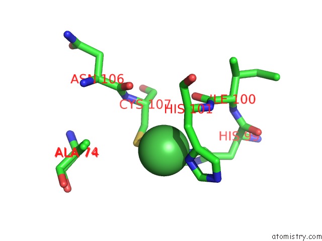
Mono view
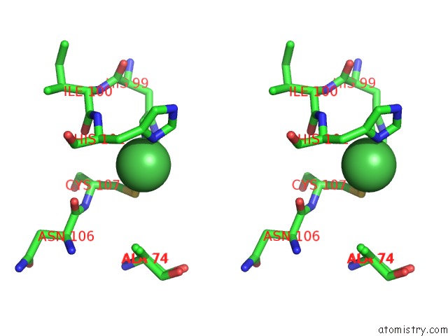
Stereo pair view
Mono view
Stereo pair view
A full contact list of Nickel with other atoms in the Ni binding
site number 2 of Crystal Structure of H74A Mutant of Helicobacter Pylori Nikr within 5.0Å range:
|
Reference:
A.L.West,
S.E.Evans,
J.M.Gonzalez,
L.G.Carter,
H.Tsuruta,
E.Pozharski,
S.L.Michel.
Ni(II) Coordination to Mixed Sites Modulates Dna Binding of Hpnikr Via A Long-Range Effect. Proc.Natl.Acad.Sci.Usa V. 109 5633 2012.
ISSN: ISSN 0027-8424
PubMed: 22451934
DOI: 10.1073/PNAS.1120283109
Page generated: Wed Oct 9 17:41:37 2024
ISSN: ISSN 0027-8424
PubMed: 22451934
DOI: 10.1073/PNAS.1120283109
Last articles
Zn in 9J0NZn in 9J0O
Zn in 9J0P
Zn in 9FJX
Zn in 9EKB
Zn in 9C0F
Zn in 9CAH
Zn in 9CH0
Zn in 9CH3
Zn in 9CH1