Nickel »
PDB 473d-4cxy »
4a2c »
Nickel in PDB 4a2c: Crystal Structure of Galactitol-1-Phosphate Dehydrogenase From Escherichia Coli
Enzymatic activity of Crystal Structure of Galactitol-1-Phosphate Dehydrogenase From Escherichia Coli
All present enzymatic activity of Crystal Structure of Galactitol-1-Phosphate Dehydrogenase From Escherichia Coli:
1.1.1.251;
1.1.1.251;
Protein crystallography data
The structure of Crystal Structure of Galactitol-1-Phosphate Dehydrogenase From Escherichia Coli, PDB code: 4a2c
was solved by
Y.Alvarez,
M.Esteban-Torres,
I.Acebron,
B.De Las Rivas,
R.Munoz,
J.M.Mancheno,
with X-Ray Crystallography technique. A brief refinement statistics is given in the table below:
| Resolution Low / High (Å) | 28.952 / 1.87 |
| Space group | P 1 21 1 |
| Cell size a, b, c (Å), α, β, γ (°) | 43.720, 77.140, 107.510, 90.00, 95.36, 90.00 |
| R / Rfree (%) | 21.27 / 27.29 |
Other elements in 4a2c:
The structure of Crystal Structure of Galactitol-1-Phosphate Dehydrogenase From Escherichia Coli also contains other interesting chemical elements:
| Zinc | (Zn) | 2 atoms |
Nickel Binding Sites:
The binding sites of Nickel atom in the Crystal Structure of Galactitol-1-Phosphate Dehydrogenase From Escherichia Coli
(pdb code 4a2c). This binding sites where shown within
5.0 Angstroms radius around Nickel atom.
In total 3 binding sites of Nickel where determined in the Crystal Structure of Galactitol-1-Phosphate Dehydrogenase From Escherichia Coli, PDB code: 4a2c:
Jump to Nickel binding site number: 1; 2; 3;
In total 3 binding sites of Nickel where determined in the Crystal Structure of Galactitol-1-Phosphate Dehydrogenase From Escherichia Coli, PDB code: 4a2c:
Jump to Nickel binding site number: 1; 2; 3;
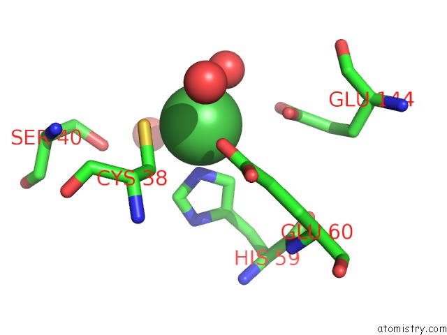
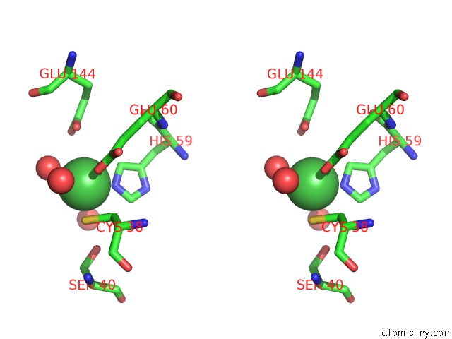
Nickel binding site 1 out of 3 in 4a2c
Go back to
Nickel binding site 1 out
of 3 in the Crystal Structure of Galactitol-1-Phosphate Dehydrogenase From Escherichia Coli
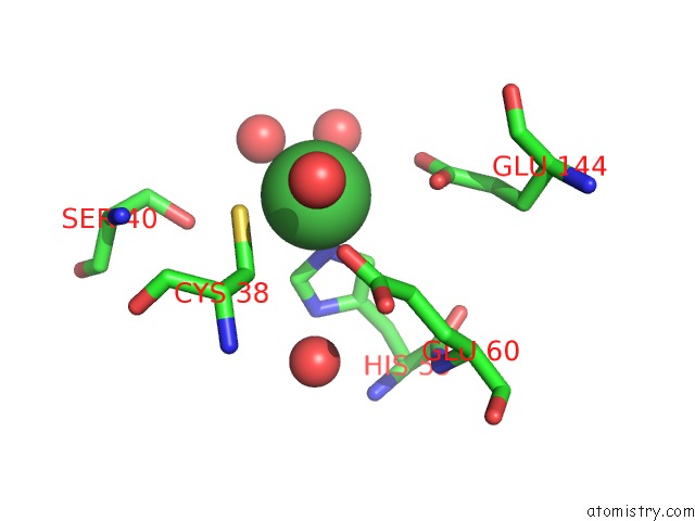
Mono view
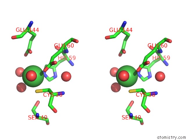
Stereo pair view
Mono view
Stereo pair view
A full contact list of Nickel with other atoms in the Ni binding
site number 1 of Crystal Structure of Galactitol-1-Phosphate Dehydrogenase From Escherichia Coli within 5.0Å range:
|
Nickel binding site 2 out of 3 in 4a2c
Go back to
Nickel binding site 2 out
of 3 in the Crystal Structure of Galactitol-1-Phosphate Dehydrogenase From Escherichia Coli
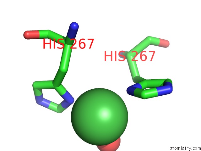
Mono view
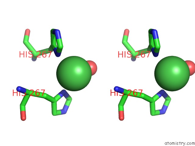
Stereo pair view
Mono view
Stereo pair view
A full contact list of Nickel with other atoms in the Ni binding
site number 2 of Crystal Structure of Galactitol-1-Phosphate Dehydrogenase From Escherichia Coli within 5.0Å range:
|
Nickel binding site 3 out of 3 in 4a2c
Go back to
Nickel binding site 3 out
of 3 in the Crystal Structure of Galactitol-1-Phosphate Dehydrogenase From Escherichia Coli
Mono view
Stereo pair view
Mono view
Stereo pair view
A full contact list of Nickel with other atoms in the Ni binding
site number 3 of Crystal Structure of Galactitol-1-Phosphate Dehydrogenase From Escherichia Coli within 5.0Å range:
|
Reference:
M.Esteban-Torres,
Y.Alvarez,
I.Acebron,
B.De Las Rivas,
R.Munoz,
G.-W.Kohring,
A.M.Roa,
M.Sobrino,
J.M.Mancheno.
The Crystal Structure of Galactitol-1-Phosphate 5-Dehydrogenase From Escherichia Coli K12 Provides Insights Into Its Anomalous Behavior on Imac Processes Febs Lett. V. 586 3127 2012.
ISSN: ISSN
PubMed: 22979983
DOI: 10.1016/J.FEBSLET.2012.07.073
Page generated: Wed Oct 9 17:58:58 2024
ISSN: ISSN
PubMed: 22979983
DOI: 10.1016/J.FEBSLET.2012.07.073
Last articles
Zn in 9MJ5Zn in 9HNW
Zn in 9G0L
Zn in 9FNE
Zn in 9DZN
Zn in 9E0I
Zn in 9D32
Zn in 9DAK
Zn in 8ZXC
Zn in 8ZUF