Nickel »
PDB 473d-4cxy »
4bu2 »
Nickel in PDB 4bu2: 60S Ribosomal Protein L27A Histidine Hydroxylase (MINA53) in Complex with Ni(II) and 2-Oxoglutarate (2OG)
Protein crystallography data
The structure of 60S Ribosomal Protein L27A Histidine Hydroxylase (MINA53) in Complex with Ni(II) and 2-Oxoglutarate (2OG), PDB code: 4bu2
was solved by
R.Chowdhury,
I.J.Clifton,
M.A.Mcdonough,
S.S.Ng,
E.Pilka,
U.Oppermann,
C.J.Schofield,
with X-Ray Crystallography technique. A brief refinement statistics is given in the table below:
| Resolution Low / High (Å) | 37.69 / 2.78 |
| Space group | P 43 3 2 |
| Cell size a, b, c (Å), α, β, γ (°) | 184.632, 184.632, 184.632, 90.00, 90.00, 90.00 |
| R / Rfree (%) | 22.8 / 23.3 |
Nickel Binding Sites:
The binding sites of Nickel atom in the 60S Ribosomal Protein L27A Histidine Hydroxylase (MINA53) in Complex with Ni(II) and 2-Oxoglutarate (2OG)
(pdb code 4bu2). This binding sites where shown within
5.0 Angstroms radius around Nickel atom.
In total 2 binding sites of Nickel where determined in the 60S Ribosomal Protein L27A Histidine Hydroxylase (MINA53) in Complex with Ni(II) and 2-Oxoglutarate (2OG), PDB code: 4bu2:
Jump to Nickel binding site number: 1; 2;
In total 2 binding sites of Nickel where determined in the 60S Ribosomal Protein L27A Histidine Hydroxylase (MINA53) in Complex with Ni(II) and 2-Oxoglutarate (2OG), PDB code: 4bu2:
Jump to Nickel binding site number: 1; 2;
Nickel binding site 1 out of 2 in 4bu2
Go back to
Nickel binding site 1 out
of 2 in the 60S Ribosomal Protein L27A Histidine Hydroxylase (MINA53) in Complex with Ni(II) and 2-Oxoglutarate (2OG)
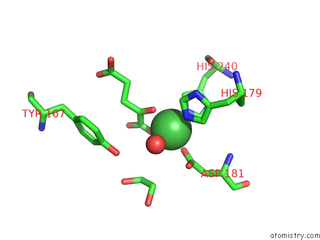
Mono view
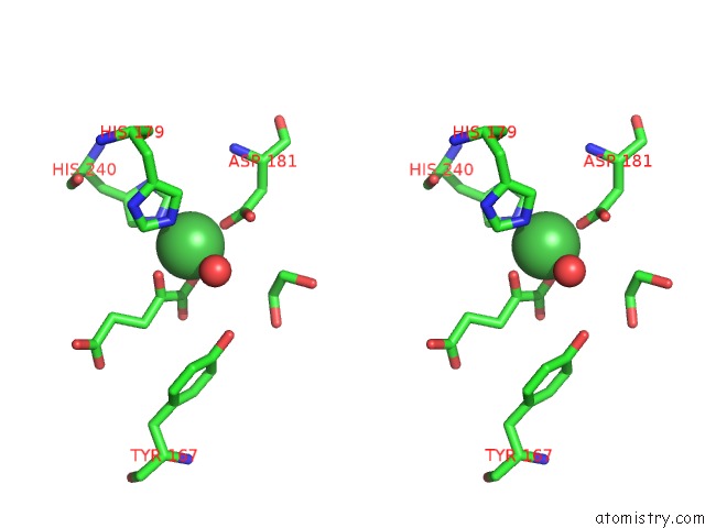
Stereo pair view
Mono view
Stereo pair view
A full contact list of Nickel with other atoms in the Ni binding
site number 1 of 60S Ribosomal Protein L27A Histidine Hydroxylase (MINA53) in Complex with Ni(II) and 2-Oxoglutarate (2OG) within 5.0Å range:
|
Nickel binding site 2 out of 2 in 4bu2
Go back to
Nickel binding site 2 out
of 2 in the 60S Ribosomal Protein L27A Histidine Hydroxylase (MINA53) in Complex with Ni(II) and 2-Oxoglutarate (2OG)
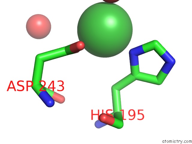
Mono view
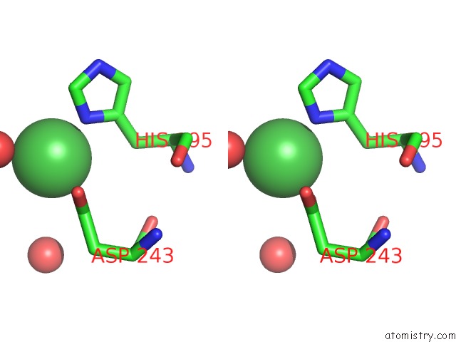
Stereo pair view
Mono view
Stereo pair view
A full contact list of Nickel with other atoms in the Ni binding
site number 2 of 60S Ribosomal Protein L27A Histidine Hydroxylase (MINA53) in Complex with Ni(II) and 2-Oxoglutarate (2OG) within 5.0Å range:
|
Reference:
R.Chowdhury,
R.Sekirnik,
N.C.Brissett,
T.Krojer,
C.-H.Ho,
S.S.Ng,
I.J.Clifton,
W.Ge,
N.J.Kershaw,
G.C.Fox,
J.R.C.Muniz,
M.Vollmar,
C.Phillips,
E.S.Pilka,
K.L.Kavanagh,
F.Von Deflt,
U.Oppermann,
M.A.Mcdonough,
A.J.Doherty,
C.J.Schofield.
Ribosomal Oxygenases Are Structurally Conserved From Prokaryotes to Humans. Nature V. 510 422 2014.
ISSN: ISSN 0028-0836
PubMed: 24814345
DOI: 10.1038/NATURE13263
Page generated: Wed Oct 9 18:02:07 2024
ISSN: ISSN 0028-0836
PubMed: 24814345
DOI: 10.1038/NATURE13263
Last articles
Zn in 9J0NZn in 9J0O
Zn in 9J0P
Zn in 9FJX
Zn in 9EKB
Zn in 9C0F
Zn in 9CAH
Zn in 9CH0
Zn in 9CH3
Zn in 9CH1