Nickel »
PDB 4d00-4gu7 »
4dun »
Nickel in PDB 4dun: 1.76A X-Ray Crystal Structure of A Putative Phenazine Biosynthesis Phzc/Phzf Protein From Clostridium Difficile (Strain 630)
Protein crystallography data
The structure of 1.76A X-Ray Crystal Structure of A Putative Phenazine Biosynthesis Phzc/Phzf Protein From Clostridium Difficile (Strain 630), PDB code: 4dun
was solved by
J.S.Brunzelle,
W.Wawrzak,
M.Kudritska,
W.F.Anderson,
A.Savchenko,
Centerfor Structural Genomics Of Infectious Diseases (Csgid),
with X-Ray Crystallography technique. A brief refinement statistics is given in the table below:
| Resolution Low / High (Å) | 23.12 / 1.76 |
| Space group | P 31 2 1 |
| Cell size a, b, c (Å), α, β, γ (°) | 63.650, 63.650, 135.120, 90.00, 90.00, 120.00 |
| R / Rfree (%) | 15.8 / 19 |
Nickel Binding Sites:
The binding sites of Nickel atom in the 1.76A X-Ray Crystal Structure of A Putative Phenazine Biosynthesis Phzc/Phzf Protein From Clostridium Difficile (Strain 630)
(pdb code 4dun). This binding sites where shown within
5.0 Angstroms radius around Nickel atom.
In total only one binding site of Nickel was determined in the 1.76A X-Ray Crystal Structure of A Putative Phenazine Biosynthesis Phzc/Phzf Protein From Clostridium Difficile (Strain 630), PDB code: 4dun:
In total only one binding site of Nickel was determined in the 1.76A X-Ray Crystal Structure of A Putative Phenazine Biosynthesis Phzc/Phzf Protein From Clostridium Difficile (Strain 630), PDB code: 4dun:
Nickel binding site 1 out of 1 in 4dun
Go back to
Nickel binding site 1 out
of 1 in the 1.76A X-Ray Crystal Structure of A Putative Phenazine Biosynthesis Phzc/Phzf Protein From Clostridium Difficile (Strain 630)
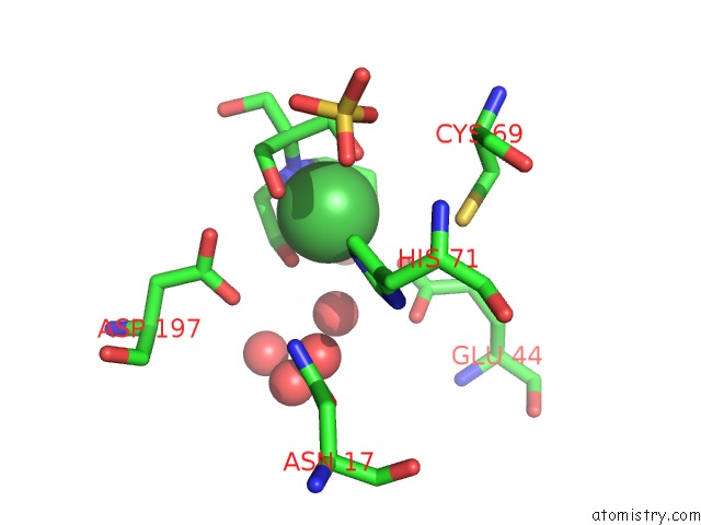
Mono view
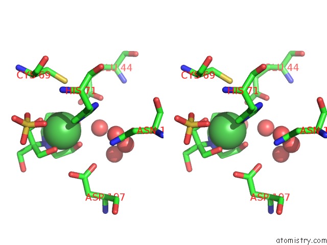
Stereo pair view
Mono view
Stereo pair view
A full contact list of Nickel with other atoms in the Ni binding
site number 1 of 1.76A X-Ray Crystal Structure of A Putative Phenazine Biosynthesis Phzc/Phzf Protein From Clostridium Difficile (Strain 630) within 5.0Å range:
|
Reference:
J.S.Brunzelle,
W.Wawrzak,
M.Kudritska,
W.F.Anderson,
A.Savchenko,
Center For Structural Genomics Of Infectiousdiseases (Csgid).
1.76A X-Ray Crystal Structure of A Putative Phenazine Biosynthesis Phzc/Phzf Protein From Clostridium Difficile (Strain 630) To Be Published.
Page generated: Mon Aug 18 19:11:14 2025
Last articles
Ni in 6ETENi in 6ETS
Ni in 6ETG
Ni in 6ELQ
Ni in 6EN9
Ni in 6EOZ
Ni in 6EN3
Ni in 6EI2
Ni in 6EK6
Ni in 6EHS