Nickel »
PDB 4d00-4gu7 »
4f9d »
Nickel in PDB 4f9d: Structure of Escherichia Coli Pgab 42-655 in Complex with Nickel
Protein crystallography data
The structure of Structure of Escherichia Coli Pgab 42-655 in Complex with Nickel, PDB code: 4f9d
was solved by
D.J.Little,
J.Poloczek,
J.C.Whitney,
H.Robinson,
M.Nitz,
P.L.Howell,
with X-Ray Crystallography technique. A brief refinement statistics is given in the table below:
| Resolution Low / High (Å) | 45.33 / 1.90 |
| Space group | P 21 21 21 |
| Cell size a, b, c (Å), α, β, γ (°) | 90.669, 102.831, 150.881, 90.00, 90.00, 90.00 |
| R / Rfree (%) | 16.8 / 19.3 |
Other elements in 4f9d:
The structure of Structure of Escherichia Coli Pgab 42-655 in Complex with Nickel also contains other interesting chemical elements:
| Calcium | (Ca) | 2 atoms |
Nickel Binding Sites:
The binding sites of Nickel atom in the Structure of Escherichia Coli Pgab 42-655 in Complex with Nickel
(pdb code 4f9d). This binding sites where shown within
5.0 Angstroms radius around Nickel atom.
In total 2 binding sites of Nickel where determined in the Structure of Escherichia Coli Pgab 42-655 in Complex with Nickel, PDB code: 4f9d:
Jump to Nickel binding site number: 1; 2;
In total 2 binding sites of Nickel where determined in the Structure of Escherichia Coli Pgab 42-655 in Complex with Nickel, PDB code: 4f9d:
Jump to Nickel binding site number: 1; 2;
Nickel binding site 1 out of 2 in 4f9d
Go back to
Nickel binding site 1 out
of 2 in the Structure of Escherichia Coli Pgab 42-655 in Complex with Nickel
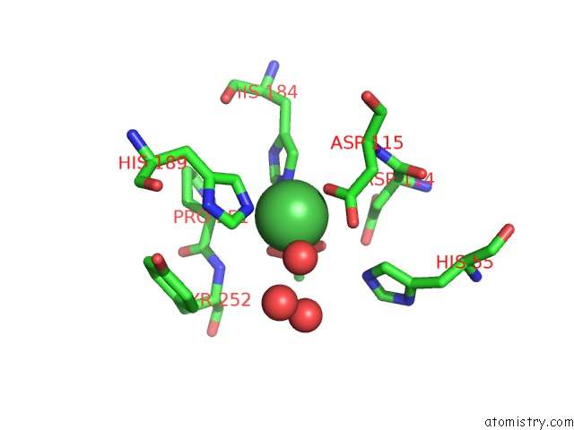
Mono view
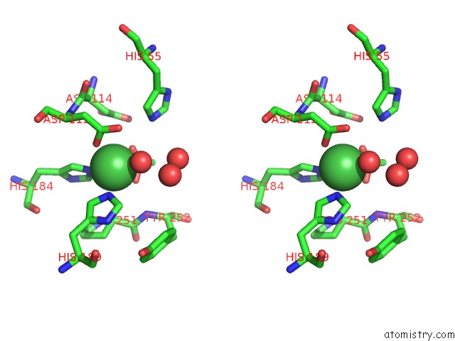
Stereo pair view
Mono view
Stereo pair view
A full contact list of Nickel with other atoms in the Ni binding
site number 1 of Structure of Escherichia Coli Pgab 42-655 in Complex with Nickel within 5.0Å range:
|
Nickel binding site 2 out of 2 in 4f9d
Go back to
Nickel binding site 2 out
of 2 in the Structure of Escherichia Coli Pgab 42-655 in Complex with Nickel
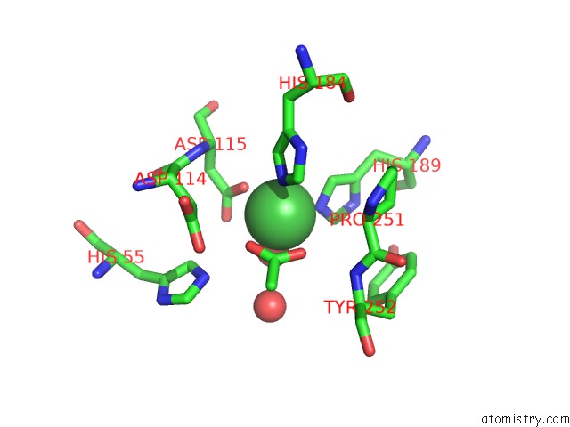
Mono view
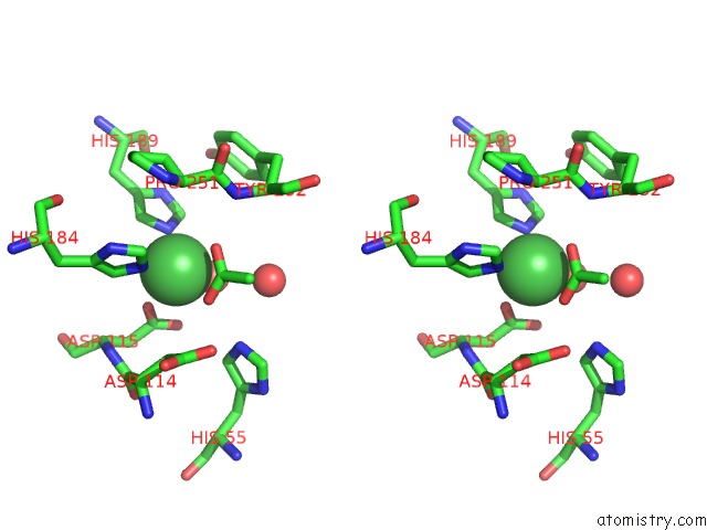
Stereo pair view
Mono view
Stereo pair view
A full contact list of Nickel with other atoms in the Ni binding
site number 2 of Structure of Escherichia Coli Pgab 42-655 in Complex with Nickel within 5.0Å range:
|
Reference:
D.J.Little,
J.Poloczek,
J.C.Whitney,
H.Robinson,
M.Nitz,
P.L.Howell.
The Structure and Metal Dependent Activity of Escherichia Coli Pgab Provides Insight Into the Partial De-N-Acetylation of Poly-B-1,6-N-Acetyl-D-Glucosamine J.Biol.Chem. V. 287 31126 2012.
ISSN: ISSN 0021-9258
PubMed: 22810235
DOI: 10.1074/JBC.M112.390005
Page generated: Wed Oct 9 18:07:44 2024
ISSN: ISSN 0021-9258
PubMed: 22810235
DOI: 10.1074/JBC.M112.390005
Last articles
Zn in 9J0NZn in 9J0O
Zn in 9J0P
Zn in 9FJX
Zn in 9EKB
Zn in 9C0F
Zn in 9CAH
Zn in 9CH0
Zn in 9CH3
Zn in 9CH1