Nickel »
PDB 4d00-4gu7 »
4gd3 »
Nickel in PDB 4gd3: Structure of E. Coli Hydrogenase-1 in Complex with Cytochrome B
Enzymatic activity of Structure of E. Coli Hydrogenase-1 in Complex with Cytochrome B
All present enzymatic activity of Structure of E. Coli Hydrogenase-1 in Complex with Cytochrome B:
1.12.99.6;
1.12.99.6;
Protein crystallography data
The structure of Structure of E. Coli Hydrogenase-1 in Complex with Cytochrome B, PDB code: 4gd3
was solved by
A.Volbeda,
J.C.Fontecilla-Camps,
C.Darnault,
with X-Ray Crystallography technique. A brief refinement statistics is given in the table below:
| Resolution Low / High (Å) | 25.00 / 3.30 |
| Space group | P 21 21 21 |
| Cell size a, b, c (Å), α, β, γ (°) | 126.000, 165.300, 212.800, 90.00, 90.00, 90.00 |
| R / Rfree (%) | 20 / 23.6 |
Other elements in 4gd3:
The structure of Structure of E. Coli Hydrogenase-1 in Complex with Cytochrome B also contains other interesting chemical elements:
| Magnesium | (Mg) | 4 atoms |
| Iron | (Fe) | 54 atoms |
| Chlorine | (Cl) | 10 atoms |
Nickel Binding Sites:
The binding sites of Nickel atom in the Structure of E. Coli Hydrogenase-1 in Complex with Cytochrome B
(pdb code 4gd3). This binding sites where shown within
5.0 Angstroms radius around Nickel atom.
In total 4 binding sites of Nickel where determined in the Structure of E. Coli Hydrogenase-1 in Complex with Cytochrome B, PDB code: 4gd3:
Jump to Nickel binding site number: 1; 2; 3; 4;
In total 4 binding sites of Nickel where determined in the Structure of E. Coli Hydrogenase-1 in Complex with Cytochrome B, PDB code: 4gd3:
Jump to Nickel binding site number: 1; 2; 3; 4;
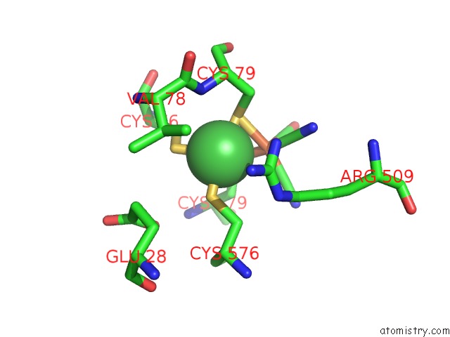
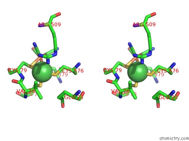
Nickel binding site 1 out of 4 in 4gd3
Go back to
Nickel binding site 1 out
of 4 in the Structure of E. Coli Hydrogenase-1 in Complex with Cytochrome B
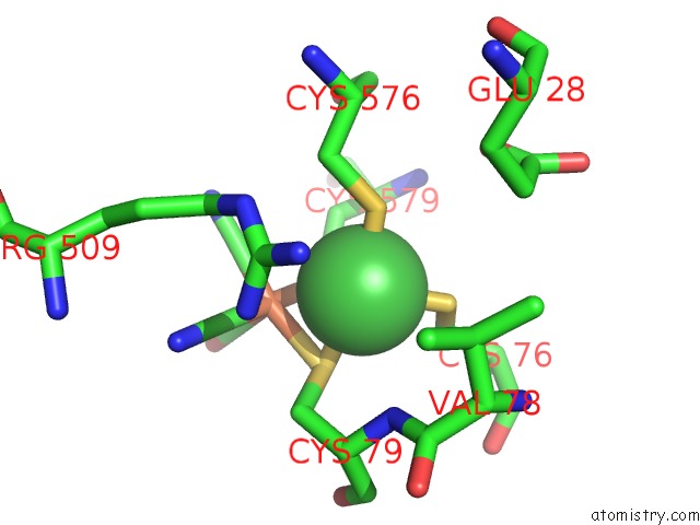
Mono view
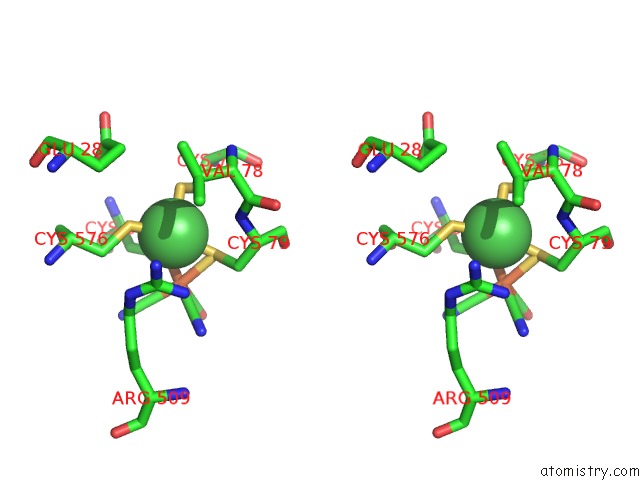
Stereo pair view
Mono view
Stereo pair view
A full contact list of Nickel with other atoms in the Ni binding
site number 1 of Structure of E. Coli Hydrogenase-1 in Complex with Cytochrome B within 5.0Å range:
|
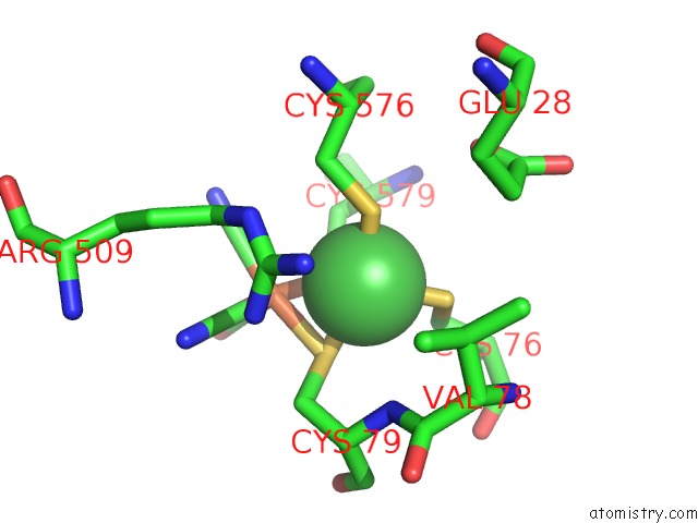
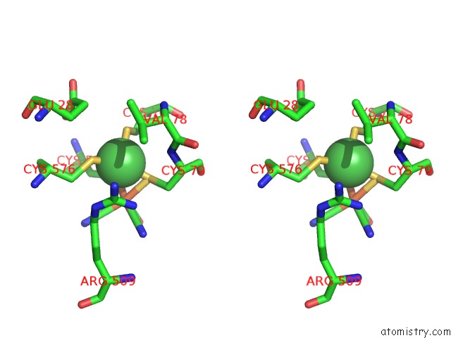
Nickel binding site 2 out of 4 in 4gd3
Go back to
Nickel binding site 2 out
of 4 in the Structure of E. Coli Hydrogenase-1 in Complex with Cytochrome B
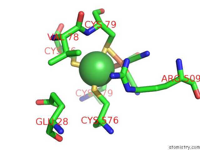
Mono view
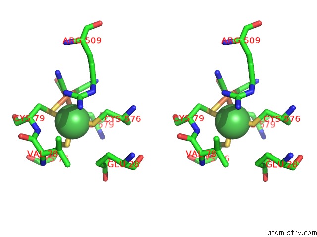
Stereo pair view
Mono view
Stereo pair view
A full contact list of Nickel with other atoms in the Ni binding
site number 2 of Structure of E. Coli Hydrogenase-1 in Complex with Cytochrome B within 5.0Å range:
|
Nickel binding site 3 out of 4 in 4gd3
Go back to
Nickel binding site 3 out
of 4 in the Structure of E. Coli Hydrogenase-1 in Complex with Cytochrome B
Mono view
Stereo pair view
Mono view
Stereo pair view
A full contact list of Nickel with other atoms in the Ni binding
site number 3 of Structure of E. Coli Hydrogenase-1 in Complex with Cytochrome B within 5.0Å range:
|
Nickel binding site 4 out of 4 in 4gd3
Go back to
Nickel binding site 4 out
of 4 in the Structure of E. Coli Hydrogenase-1 in Complex with Cytochrome B
Mono view
Stereo pair view
Mono view
Stereo pair view
A full contact list of Nickel with other atoms in the Ni binding
site number 4 of Structure of E. Coli Hydrogenase-1 in Complex with Cytochrome B within 5.0Å range:
|
Reference:
A.Volbeda,
C.Darnault,
A.Parkin,
F.Sargent,
F.A.Armstrong,
J.C.Fontecilla-Camps.
Crystal Structure of the O(2)-Tolerant Membrane-Bound Hydrogenase 1 From Escherichia Coli in Complex with Its Cognate Cytochrome B. Structure V. 21 184 2013.
ISSN: ISSN 0969-2126
PubMed: 23260654
DOI: 10.1016/J.STR.2012.11.010
Page generated: Wed Oct 9 18:08:40 2024
ISSN: ISSN 0969-2126
PubMed: 23260654
DOI: 10.1016/J.STR.2012.11.010
Last articles
Zn in 9J0NZn in 9J0O
Zn in 9J0P
Zn in 9FJX
Zn in 9EKB
Zn in 9C0F
Zn in 9CAH
Zn in 9CH0
Zn in 9CH3
Zn in 9CH1