Nickel »
PDB 4guh-4jz4 »
4i73 »
Nickel in PDB 4i73: Crystal Structure of the Trypanosoma Brucei Inosine-Adenosine- Guanosine Nucleoside Hydrolase in Complex with Compound Uamc-00312
Enzymatic activity of Crystal Structure of the Trypanosoma Brucei Inosine-Adenosine- Guanosine Nucleoside Hydrolase in Complex with Compound Uamc-00312
All present enzymatic activity of Crystal Structure of the Trypanosoma Brucei Inosine-Adenosine- Guanosine Nucleoside Hydrolase in Complex with Compound Uamc-00312:
3.2.2.1;
3.2.2.1;
Protein crystallography data
The structure of Crystal Structure of the Trypanosoma Brucei Inosine-Adenosine- Guanosine Nucleoside Hydrolase in Complex with Compound Uamc-00312, PDB code: 4i73
was solved by
F.Giannese,
M.Degano,
with X-Ray Crystallography technique. A brief refinement statistics is given in the table below:
| Resolution Low / High (Å) | 71.83 / 2.18 |
| Space group | P 1 21 1 |
| Cell size a, b, c (Å), α, β, γ (°) | 63.170, 131.670, 71.850, 90.00, 91.33, 90.00 |
| R / Rfree (%) | 20 / 24.8 |
Other elements in 4i73:
The structure of Crystal Structure of the Trypanosoma Brucei Inosine-Adenosine- Guanosine Nucleoside Hydrolase in Complex with Compound Uamc-00312 also contains other interesting chemical elements:
| Calcium | (Ca) | 4 atoms |
Nickel Binding Sites:
Pages:
>>> Page 1 <<< Page 2, Binding sites: 11 - 17;Binding sites:
The binding sites of Nickel atom in the Crystal Structure of the Trypanosoma Brucei Inosine-Adenosine- Guanosine Nucleoside Hydrolase in Complex with Compound Uamc-00312 (pdb code 4i73). This binding sites where shown within 5.0 Angstroms radius around Nickel atom.In total 17 binding sites of Nickel where determined in the Crystal Structure of the Trypanosoma Brucei Inosine-Adenosine- Guanosine Nucleoside Hydrolase in Complex with Compound Uamc-00312, PDB code: 4i73:
Jump to Nickel binding site number: 1; 2; 3; 4; 5; 6; 7; 8; 9; 10;
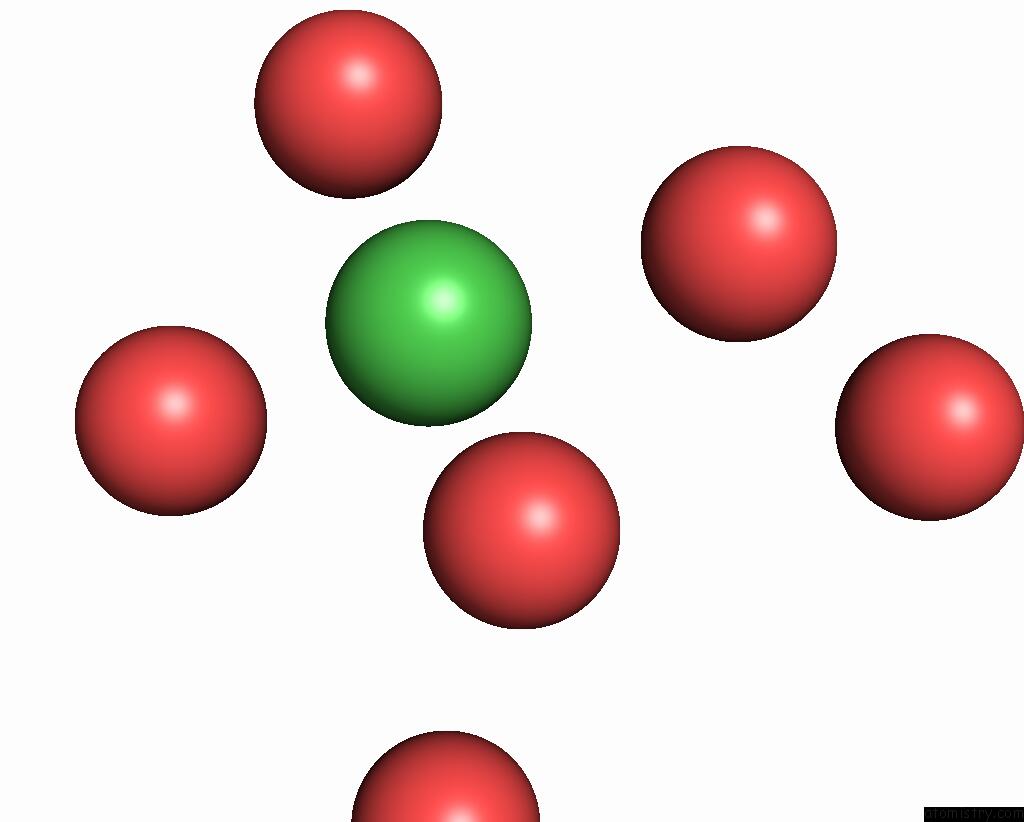
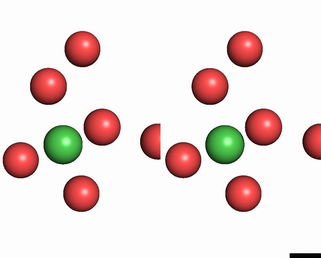
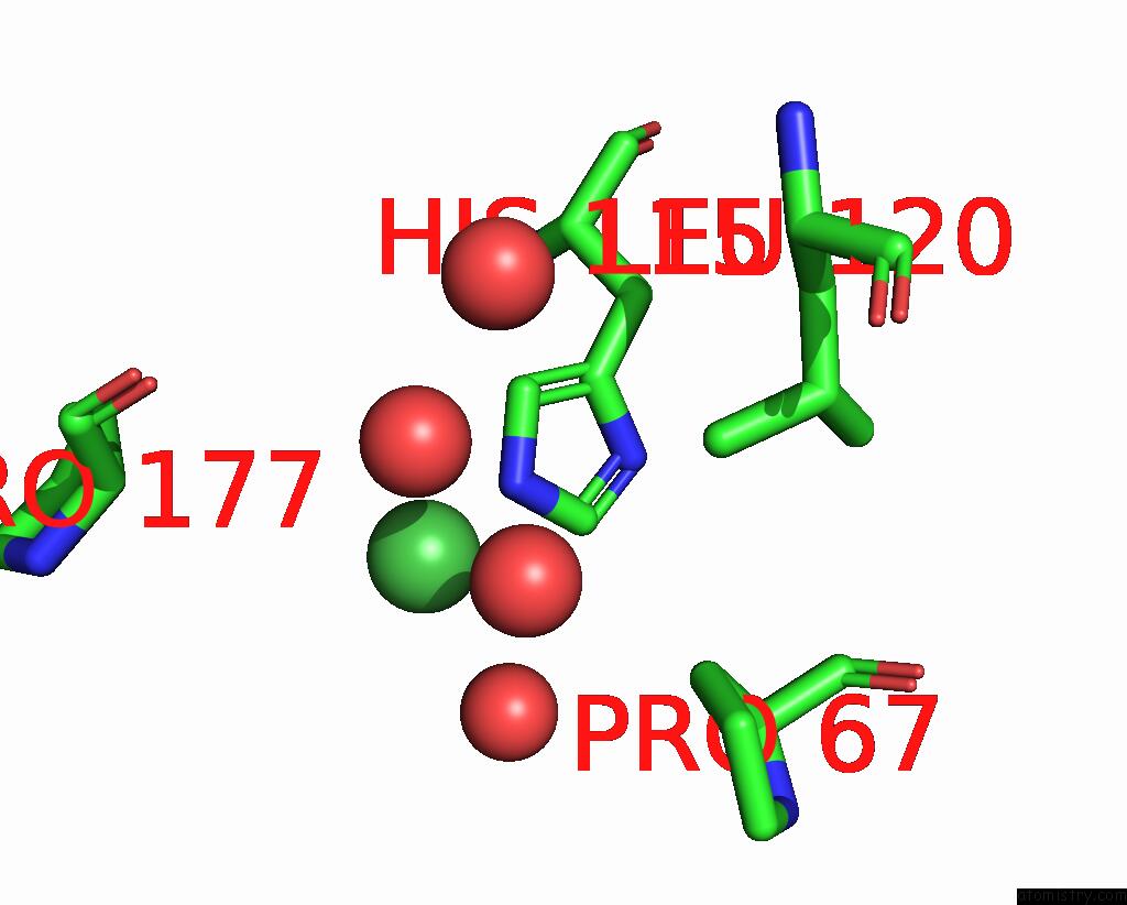
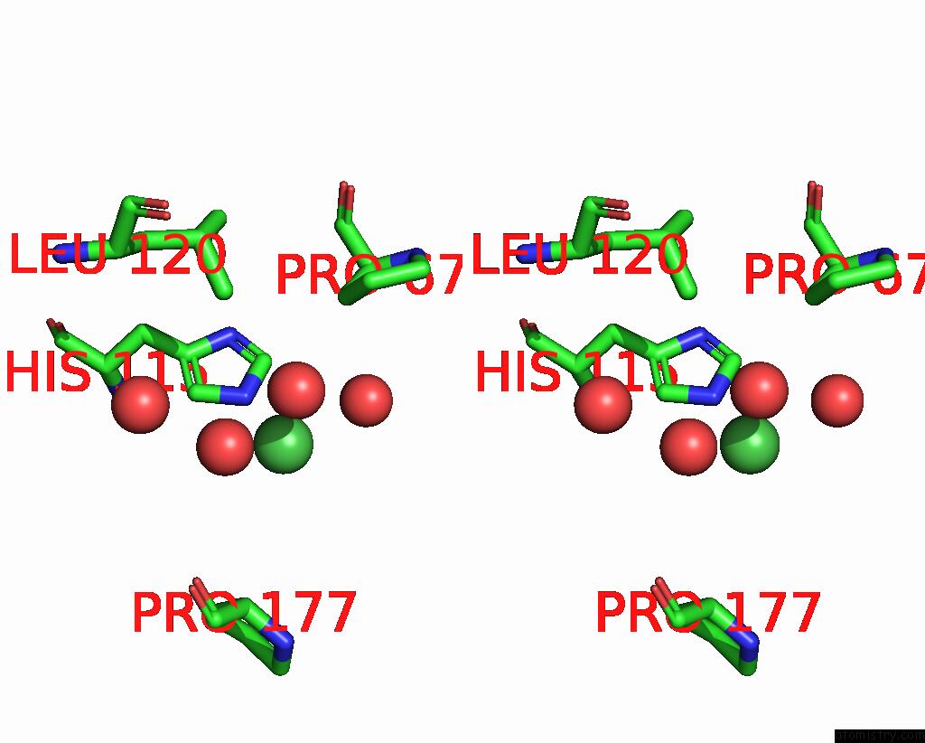
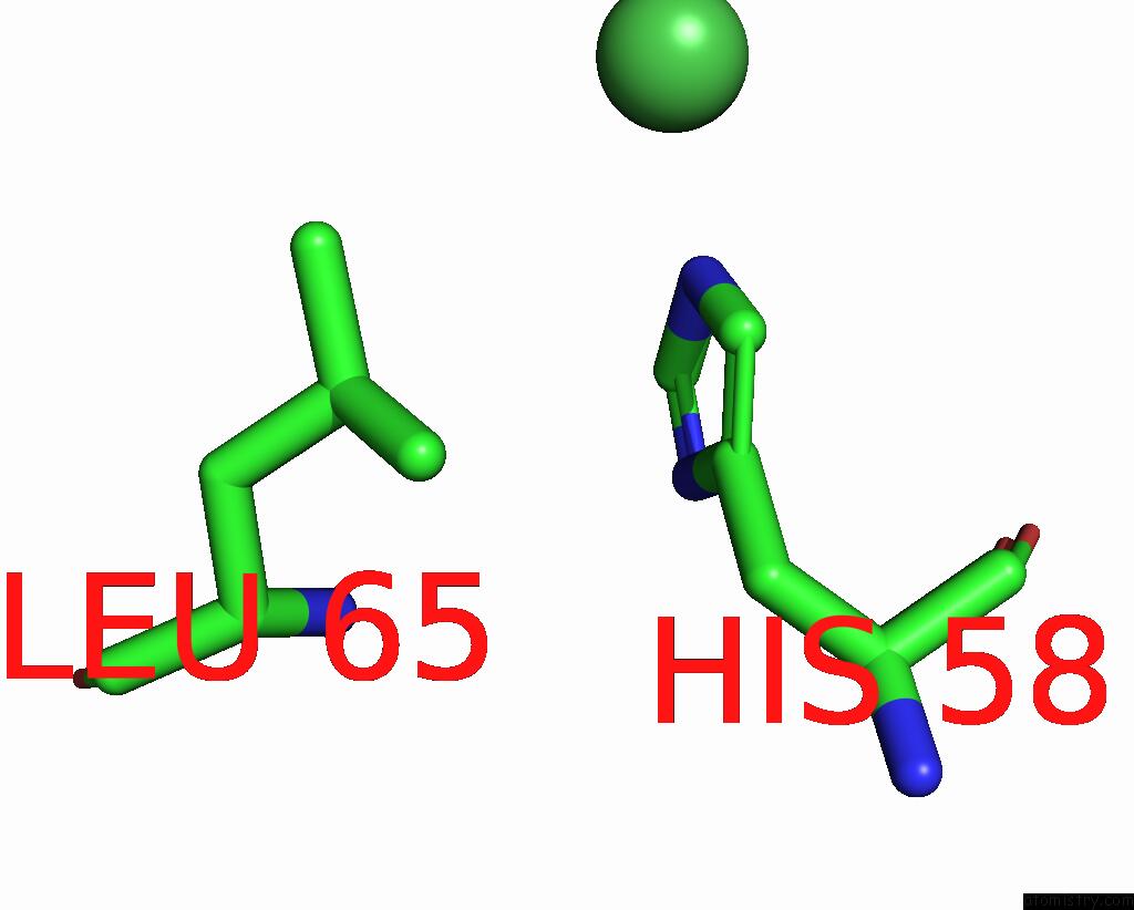
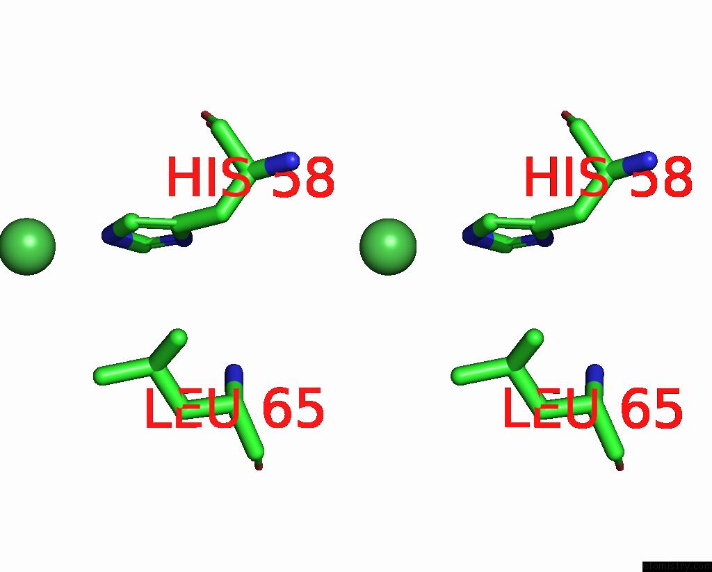
Nickel binding site 1 out of 17 in 4i73
Go back to
Nickel binding site 1 out
of 17 in the Crystal Structure of the Trypanosoma Brucei Inosine-Adenosine- Guanosine Nucleoside Hydrolase in Complex with Compound Uamc-00312
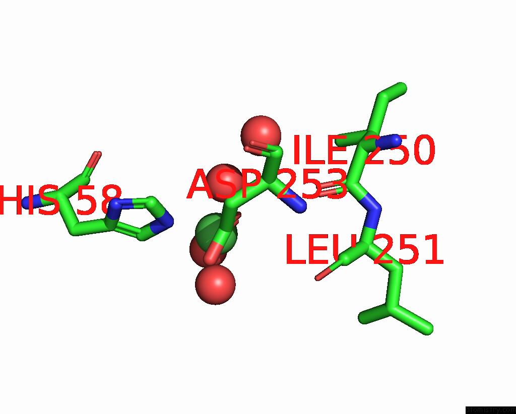
Mono view
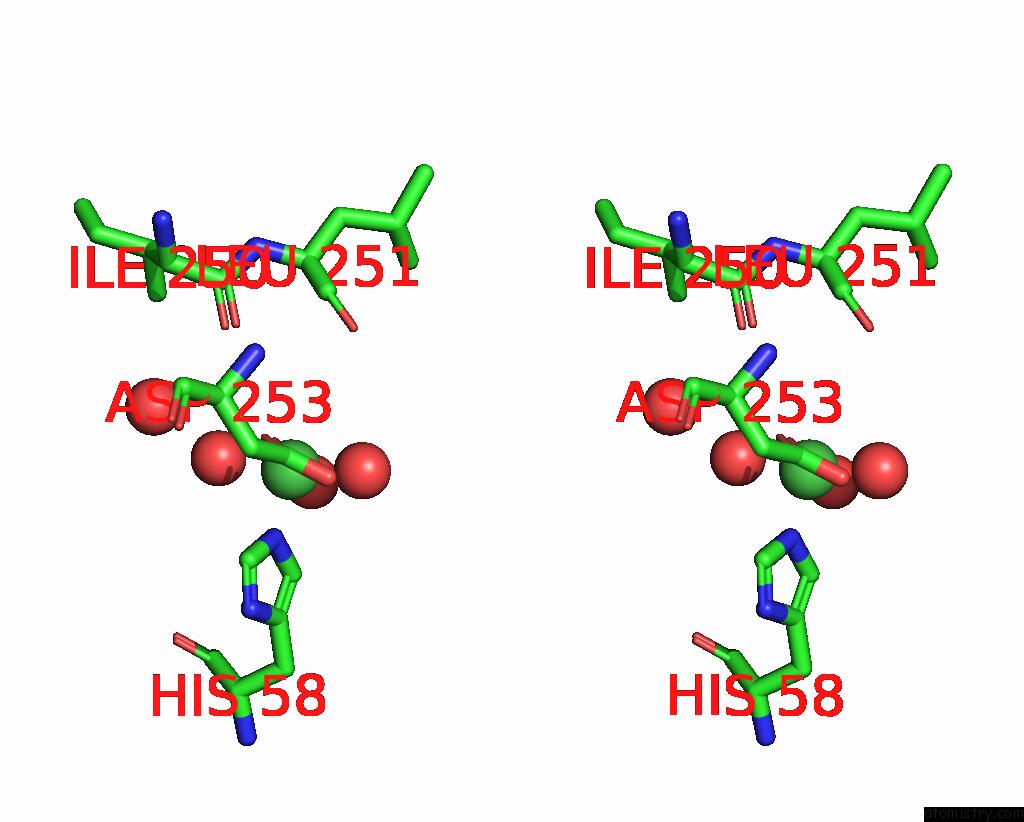
Stereo pair view
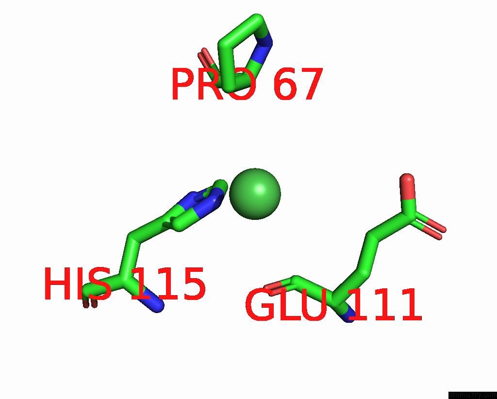
Mono view
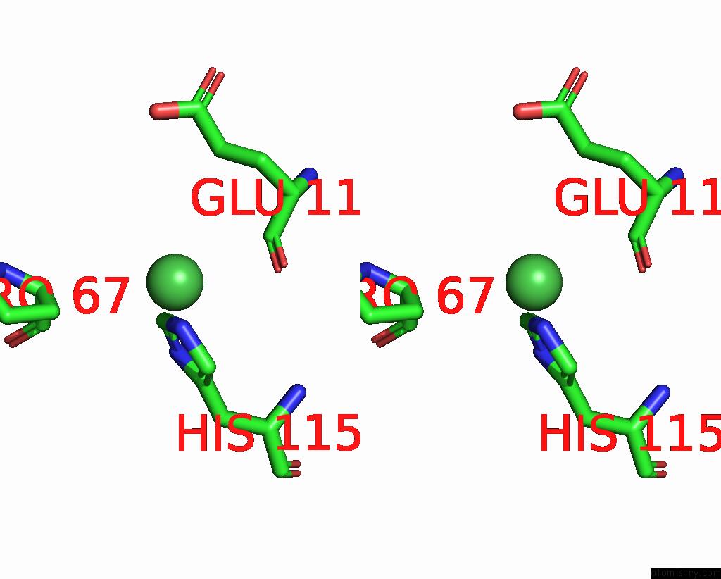
Stereo pair view
A full contact list of Nickel with other atoms in the Ni binding
site number 1 of Crystal Structure of the Trypanosoma Brucei Inosine-Adenosine- Guanosine Nucleoside Hydrolase in Complex with Compound Uamc-00312 within 5.0Å range:
|
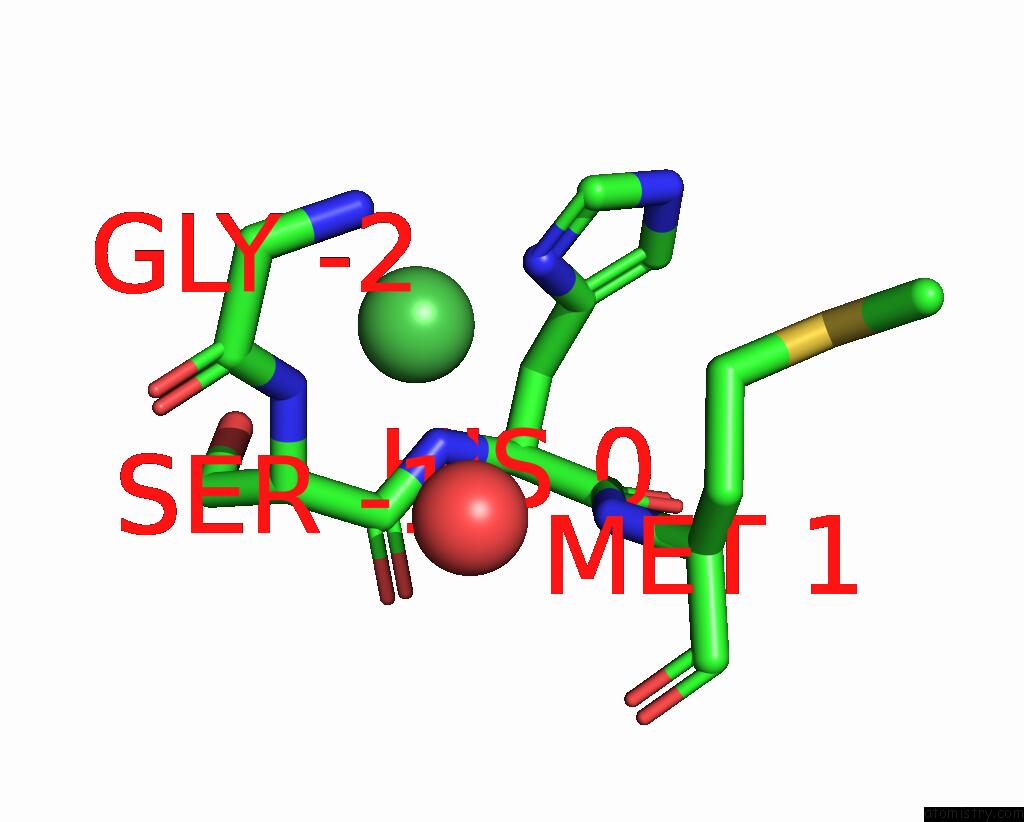
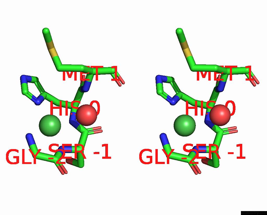
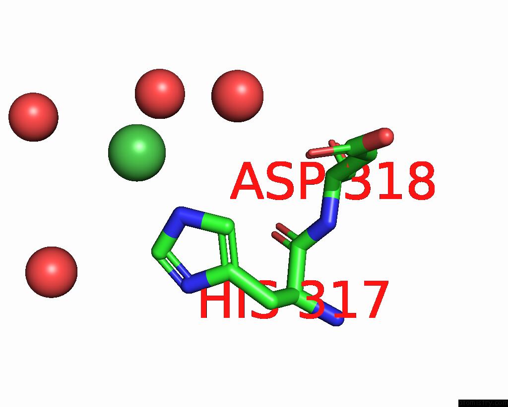
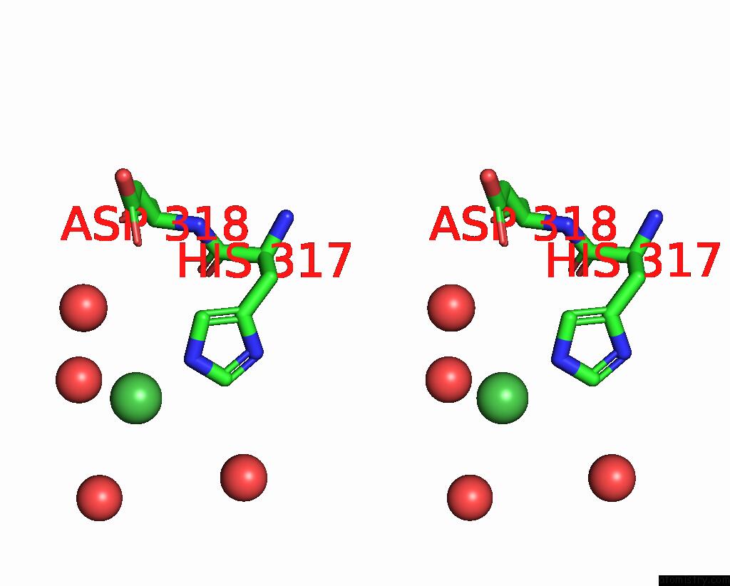
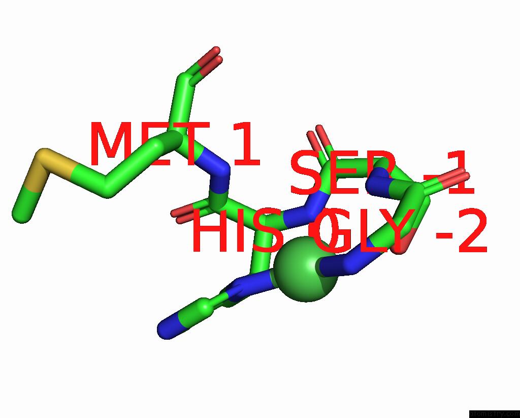
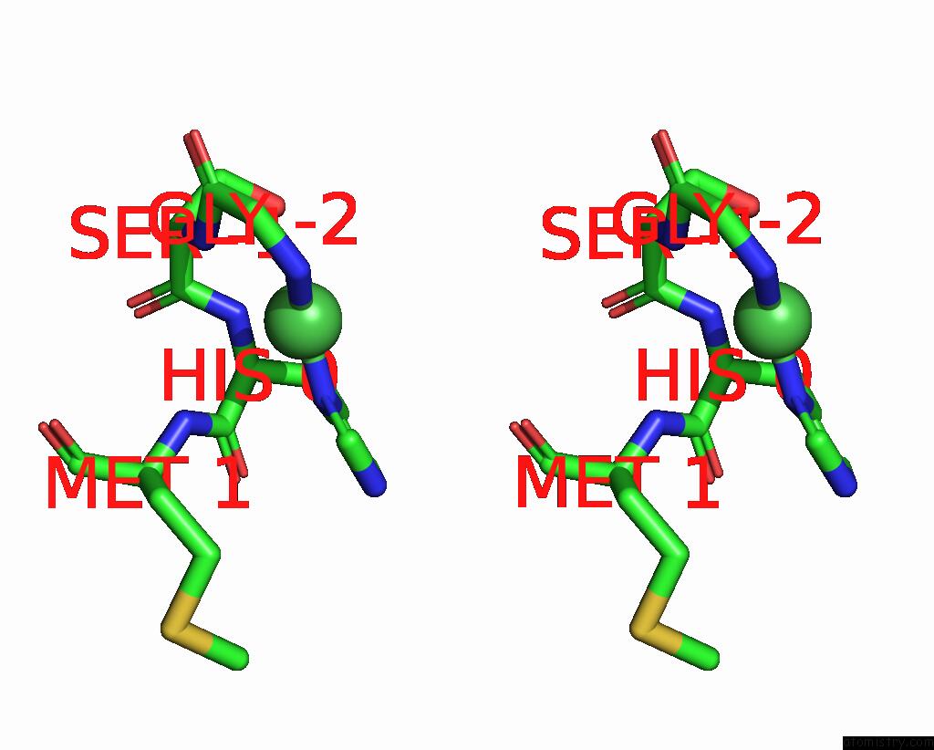
Nickel binding site 2 out of 17 in 4i73
Go back to
Nickel binding site 2 out
of 17 in the Crystal Structure of the Trypanosoma Brucei Inosine-Adenosine- Guanosine Nucleoside Hydrolase in Complex with Compound Uamc-00312
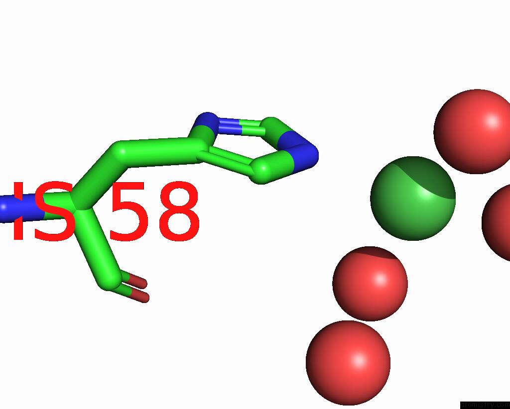
Mono view
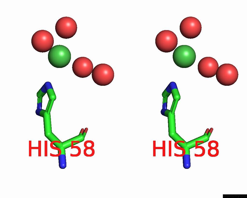
Stereo pair view
Mono view
Stereo pair view
A full contact list of Nickel with other atoms in the Ni binding
site number 2 of Crystal Structure of the Trypanosoma Brucei Inosine-Adenosine- Guanosine Nucleoside Hydrolase in Complex with Compound Uamc-00312 within 5.0Å range:
|
Nickel binding site 3 out of 17 in 4i73
Go back to
Nickel binding site 3 out
of 17 in the Crystal Structure of the Trypanosoma Brucei Inosine-Adenosine- Guanosine Nucleoside Hydrolase in Complex with Compound Uamc-00312
Mono view
Stereo pair view
Mono view
Stereo pair view
A full contact list of Nickel with other atoms in the Ni binding
site number 3 of Crystal Structure of the Trypanosoma Brucei Inosine-Adenosine- Guanosine Nucleoside Hydrolase in Complex with Compound Uamc-00312 within 5.0Å range:
|
Nickel binding site 4 out of 17 in 4i73
Go back to
Nickel binding site 4 out
of 17 in the Crystal Structure of the Trypanosoma Brucei Inosine-Adenosine- Guanosine Nucleoside Hydrolase in Complex with Compound Uamc-00312
Mono view
Stereo pair view
Mono view
Stereo pair view
A full contact list of Nickel with other atoms in the Ni binding
site number 4 of Crystal Structure of the Trypanosoma Brucei Inosine-Adenosine- Guanosine Nucleoside Hydrolase in Complex with Compound Uamc-00312 within 5.0Å range:
|
Nickel binding site 5 out of 17 in 4i73
Go back to
Nickel binding site 5 out
of 17 in the Crystal Structure of the Trypanosoma Brucei Inosine-Adenosine- Guanosine Nucleoside Hydrolase in Complex with Compound Uamc-00312
Mono view
Stereo pair view
Mono view
Stereo pair view
| A full contact list of Nickel with other atoms in the Ni binding site number 5 of Crystal Structure of the Trypanosoma Brucei Inosine-Adenosine- Guanosine Nucleoside Hydrolase in Complex with Compound Uamc-00312 within 5.0Å range: |
Nickel binding site 6 out of 17 in 4i73
Go back to
Nickel binding site 6 out
of 17 in the Crystal Structure of the Trypanosoma Brucei Inosine-Adenosine- Guanosine Nucleoside Hydrolase in Complex with Compound Uamc-00312
Mono view
Stereo pair view
Mono view
Stereo pair view
A full contact list of Nickel with other atoms in the Ni binding
site number 6 of Crystal Structure of the Trypanosoma Brucei Inosine-Adenosine- Guanosine Nucleoside Hydrolase in Complex with Compound Uamc-00312 within 5.0Å range:
|
Nickel binding site 7 out of 17 in 4i73
Go back to
Nickel binding site 7 out
of 17 in the Crystal Structure of the Trypanosoma Brucei Inosine-Adenosine- Guanosine Nucleoside Hydrolase in Complex with Compound Uamc-00312
Mono view
Stereo pair view
Mono view
Stereo pair view
A full contact list of Nickel with other atoms in the Ni binding
site number 7 of Crystal Structure of the Trypanosoma Brucei Inosine-Adenosine- Guanosine Nucleoside Hydrolase in Complex with Compound Uamc-00312 within 5.0Å range:
|
Nickel binding site 8 out of 17 in 4i73
Go back to
Nickel binding site 8 out
of 17 in the Crystal Structure of the Trypanosoma Brucei Inosine-Adenosine- Guanosine Nucleoside Hydrolase in Complex with Compound Uamc-00312
Mono view
Stereo pair view
Mono view
Stereo pair view
A full contact list of Nickel with other atoms in the Ni binding
site number 8 of Crystal Structure of the Trypanosoma Brucei Inosine-Adenosine- Guanosine Nucleoside Hydrolase in Complex with Compound Uamc-00312 within 5.0Å range:
|
Nickel binding site 9 out of 17 in 4i73
Go back to
Nickel binding site 9 out
of 17 in the Crystal Structure of the Trypanosoma Brucei Inosine-Adenosine- Guanosine Nucleoside Hydrolase in Complex with Compound Uamc-00312
Mono view
Stereo pair view
Mono view
Stereo pair view
A full contact list of Nickel with other atoms in the Ni binding
site number 9 of Crystal Structure of the Trypanosoma Brucei Inosine-Adenosine- Guanosine Nucleoside Hydrolase in Complex with Compound Uamc-00312 within 5.0Å range:
|
Nickel binding site 10 out of 17 in 4i73
Go back to
Nickel binding site 10 out
of 17 in the Crystal Structure of the Trypanosoma Brucei Inosine-Adenosine- Guanosine Nucleoside Hydrolase in Complex with Compound Uamc-00312
Mono view
Stereo pair view
Mono view
Stereo pair view
A full contact list of Nickel with other atoms in the Ni binding
site number 10 of Crystal Structure of the Trypanosoma Brucei Inosine-Adenosine- Guanosine Nucleoside Hydrolase in Complex with Compound Uamc-00312 within 5.0Å range:
|
Reference:
F.Giannese,
M.Berg,
P.Van Der Veken,
V.Castagna,
P.Tornaghi,
K.Augustyns,
M.Degano.
Structures of Purine Nucleosidase From Trypanosoma Brucei Bound to Isozyme-Specific Trypanocidals and A Novel Metalorganic Inhibitor Acta Crystallogr.,Sect.D V. 69 1553 2013.
ISSN: ISSN 0907-4449
PubMed: 23897478
DOI: 10.1107/S0907444913010792
Page generated: Wed Oct 9 18:12:43 2024
ISSN: ISSN 0907-4449
PubMed: 23897478
DOI: 10.1107/S0907444913010792
Last articles
Zn in 9J0NZn in 9J0O
Zn in 9J0P
Zn in 9FJX
Zn in 9EKB
Zn in 9C0F
Zn in 9CAH
Zn in 9CH0
Zn in 9CH3
Zn in 9CH1