Nickel »
PDB 4lto-4ofl »
4m4m »
Nickel in PDB 4m4m: The Structure of Ni T6 Bovine Insulin
Protein crystallography data
The structure of The Structure of Ni T6 Bovine Insulin, PDB code: 4m4m
was solved by
C.G.Frankaer,
P.Harris,
K.Stahl,
with X-Ray Crystallography technique. A brief refinement statistics is given in the table below:
| Resolution Low / High (Å) | 16.78 / 1.50 |
| Space group | H 3 |
| Cell size a, b, c (Å), α, β, γ (°) | 80.850, 80.850, 33.360, 90.00, 90.00, 120.00 |
| R / Rfree (%) | 18.2 / 22.4 |
Nickel Binding Sites:
The binding sites of Nickel atom in the The Structure of Ni T6 Bovine Insulin
(pdb code 4m4m). This binding sites where shown within
5.0 Angstroms radius around Nickel atom.
In total 2 binding sites of Nickel where determined in the The Structure of Ni T6 Bovine Insulin, PDB code: 4m4m:
Jump to Nickel binding site number: 1; 2;
In total 2 binding sites of Nickel where determined in the The Structure of Ni T6 Bovine Insulin, PDB code: 4m4m:
Jump to Nickel binding site number: 1; 2;
Nickel binding site 1 out of 2 in 4m4m
Go back to
Nickel binding site 1 out
of 2 in the The Structure of Ni T6 Bovine Insulin

Mono view
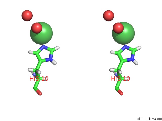
Stereo pair view
Mono view
Stereo pair view
A full contact list of Nickel with other atoms in the Ni binding
site number 1 of The Structure of Ni T6 Bovine Insulin within 5.0Å range:
|
Nickel binding site 2 out of 2 in 4m4m
Go back to
Nickel binding site 2 out
of 2 in the The Structure of Ni T6 Bovine Insulin

Mono view

Stereo pair view
Mono view
Stereo pair view
A full contact list of Nickel with other atoms in the Ni binding
site number 2 of The Structure of Ni T6 Bovine Insulin within 5.0Å range:
|
Reference:
C.G.Frankaer,
S.Mossin,
K.Stahl,
P.Harris.
Towards Accurate Structural Characterization of Metal Centres in Protein Crystals: the Structures of Ni and Cu T6 Bovine Insulin Derivatives. Acta Crystallogr.,Sect.D V. 70 110 2014.
ISSN: ISSN 0907-4449
PubMed: 24419384
DOI: 10.1107/S1399004713029040
Page generated: Mon Aug 18 19:29:06 2025
ISSN: ISSN 0907-4449
PubMed: 24419384
DOI: 10.1107/S1399004713029040
Last articles
Rb in 6DOTRb in 5YLV
Rb in 5AW8
Rb in 5YCK
Rb in 5AW6
Rb in 5AW4
Rb in 5AW7
Rb in 5AW5
Rb in 4E8P
Rb in 4R8C