Nickel »
PDB 4ofo-4rro »
4r8z »
Nickel in PDB 4r8z: Crystal Structure of PA4781 Hd-Gyp Domain From Pseudomonas Aeruginosa at 2.2A Resolution Showing A Bi-Metallic Ni Ion Center
Enzymatic activity of Crystal Structure of PA4781 Hd-Gyp Domain From Pseudomonas Aeruginosa at 2.2A Resolution Showing A Bi-Metallic Ni Ion Center
All present enzymatic activity of Crystal Structure of PA4781 Hd-Gyp Domain From Pseudomonas Aeruginosa at 2.2A Resolution Showing A Bi-Metallic Ni Ion Center:
3.1.4.1;
3.1.4.1;
Protein crystallography data
The structure of Crystal Structure of PA4781 Hd-Gyp Domain From Pseudomonas Aeruginosa at 2.2A Resolution Showing A Bi-Metallic Ni Ion Center, PDB code: 4r8z
was solved by
G.Giardina,
F.Cutruzzolaa,
S.Rinaldo,
V.Stelitano,
with X-Ray Crystallography technique. A brief refinement statistics is given in the table below:
| Resolution Low / High (Å) | 46.99 / 2.20 |
| Space group | P 41 |
| Cell size a, b, c (Å), α, β, γ (°) | 93.984, 93.984, 78.469, 90.00, 90.00, 90.00 |
| R / Rfree (%) | 17.7 / 21.3 |
Other elements in 4r8z:
The structure of Crystal Structure of PA4781 Hd-Gyp Domain From Pseudomonas Aeruginosa at 2.2A Resolution Showing A Bi-Metallic Ni Ion Center also contains other interesting chemical elements:
| Chlorine | (Cl) | 2 atoms |
Nickel Binding Sites:
The binding sites of Nickel atom in the Crystal Structure of PA4781 Hd-Gyp Domain From Pseudomonas Aeruginosa at 2.2A Resolution Showing A Bi-Metallic Ni Ion Center
(pdb code 4r8z). This binding sites where shown within
5.0 Angstroms radius around Nickel atom.
In total 4 binding sites of Nickel where determined in the Crystal Structure of PA4781 Hd-Gyp Domain From Pseudomonas Aeruginosa at 2.2A Resolution Showing A Bi-Metallic Ni Ion Center, PDB code: 4r8z:
Jump to Nickel binding site number: 1; 2; 3; 4;
In total 4 binding sites of Nickel where determined in the Crystal Structure of PA4781 Hd-Gyp Domain From Pseudomonas Aeruginosa at 2.2A Resolution Showing A Bi-Metallic Ni Ion Center, PDB code: 4r8z:
Jump to Nickel binding site number: 1; 2; 3; 4;
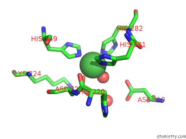
Nickel binding site 1 out of 4 in 4r8z
Go back to
Nickel binding site 1 out
of 4 in the Crystal Structure of PA4781 Hd-Gyp Domain From Pseudomonas Aeruginosa at 2.2A Resolution Showing A Bi-Metallic Ni Ion Center
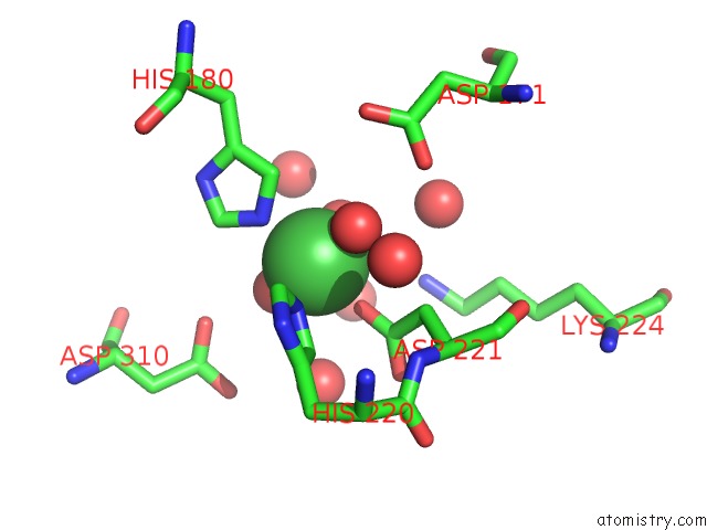
Mono view
Stereo pair view
Mono view
Stereo pair view
A full contact list of Nickel with other atoms in the Ni binding
site number 1 of Crystal Structure of PA4781 Hd-Gyp Domain From Pseudomonas Aeruginosa at 2.2A Resolution Showing A Bi-Metallic Ni Ion Center within 5.0Å range:
|
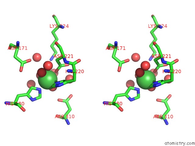
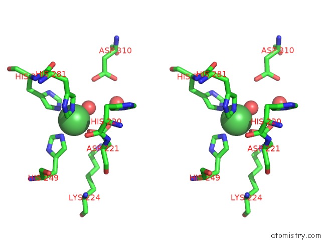
Nickel binding site 2 out of 4 in 4r8z
Go back to
Nickel binding site 2 out
of 4 in the Crystal Structure of PA4781 Hd-Gyp Domain From Pseudomonas Aeruginosa at 2.2A Resolution Showing A Bi-Metallic Ni Ion Center
Mono view
Stereo pair view
Mono view
Stereo pair view
A full contact list of Nickel with other atoms in the Ni binding
site number 2 of Crystal Structure of PA4781 Hd-Gyp Domain From Pseudomonas Aeruginosa at 2.2A Resolution Showing A Bi-Metallic Ni Ion Center within 5.0Å range:
|
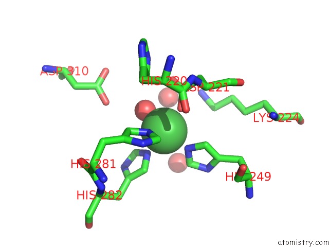
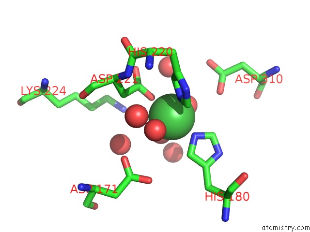
Nickel binding site 3 out of 4 in 4r8z
Go back to
Nickel binding site 3 out
of 4 in the Crystal Structure of PA4781 Hd-Gyp Domain From Pseudomonas Aeruginosa at 2.2A Resolution Showing A Bi-Metallic Ni Ion Center
Mono view
Stereo pair view
Mono view
Stereo pair view
A full contact list of Nickel with other atoms in the Ni binding
site number 3 of Crystal Structure of PA4781 Hd-Gyp Domain From Pseudomonas Aeruginosa at 2.2A Resolution Showing A Bi-Metallic Ni Ion Center within 5.0Å range:
|
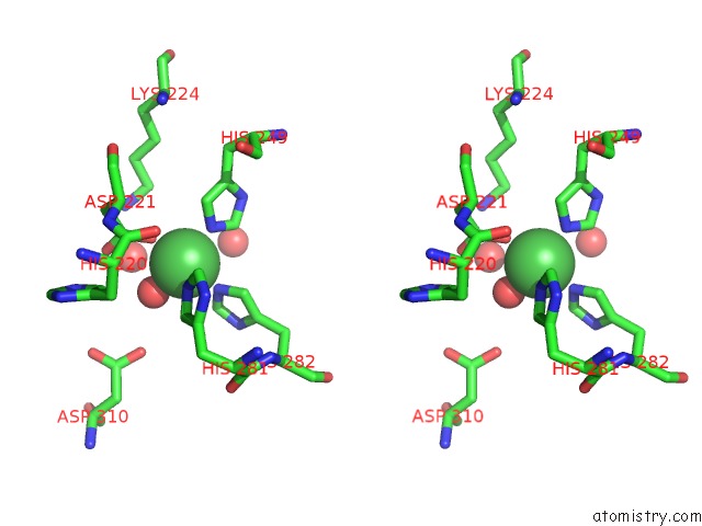
Nickel binding site 4 out of 4 in 4r8z
Go back to
Nickel binding site 4 out
of 4 in the Crystal Structure of PA4781 Hd-Gyp Domain From Pseudomonas Aeruginosa at 2.2A Resolution Showing A Bi-Metallic Ni Ion Center
Mono view
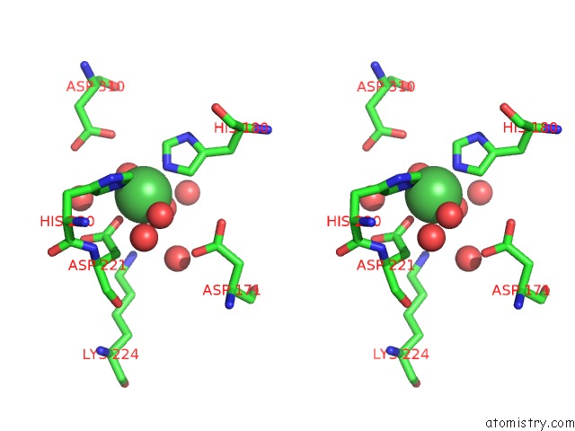
Stereo pair view
Mono view
Stereo pair view
A full contact list of Nickel with other atoms in the Ni binding
site number 4 of Crystal Structure of PA4781 Hd-Gyp Domain From Pseudomonas Aeruginosa at 2.2A Resolution Showing A Bi-Metallic Ni Ion Center within 5.0Å range:
|
Reference:
S.Rinaldo,
A.Paiardini,
V.Stelitano,
P.Brunotti,
L.Cervoni,
S.Fernicola,
C.Protano,
M.Vitali,
F.Cutruzzola,
G.Giardina.
The Structural Basis of Functional Diversification of Hd-Gyp Domain Revealed By the Pseudomonas Aeruginosa PA4781 Protein Displaying An Unselective Bi-Metallic Binding Site. J.Bacteriol. 2015.
ISSN: ESSN 1098-5530
PubMed: 25691523
DOI: 10.1128/JB.02606-14
Page generated: Wed Oct 9 18:42:14 2024
ISSN: ESSN 1098-5530
PubMed: 25691523
DOI: 10.1128/JB.02606-14
Last articles
Zn in 9J0NZn in 9J0O
Zn in 9J0P
Zn in 9FJX
Zn in 9EKB
Zn in 9C0F
Zn in 9CAH
Zn in 9CH0
Zn in 9CH3
Zn in 9CH1