Nickel »
PDB 5bu6-5e6j »
5c74 »
Nickel in PDB 5c74: Structure of A Novel Protein Arginine Methyltransferase
Protein crystallography data
The structure of Structure of A Novel Protein Arginine Methyltransferase, PDB code: 5c74
was solved by
F.Lv,
J.Ding,
with X-Ray Crystallography technique. A brief refinement statistics is given in the table below:
| Resolution Low / High (Å) | 42.16 / 1.90 |
| Space group | P 41 21 2 |
| Cell size a, b, c (Å), α, β, γ (°) | 107.570, 107.570, 87.522, 90.00, 90.00, 90.00 |
| R / Rfree (%) | 19.3 / 23.2 |
Nickel Binding Sites:
The binding sites of Nickel atom in the Structure of A Novel Protein Arginine Methyltransferase
(pdb code 5c74). This binding sites where shown within
5.0 Angstroms radius around Nickel atom.
In total 4 binding sites of Nickel where determined in the Structure of A Novel Protein Arginine Methyltransferase, PDB code: 5c74:
Jump to Nickel binding site number: 1; 2; 3; 4;
In total 4 binding sites of Nickel where determined in the Structure of A Novel Protein Arginine Methyltransferase, PDB code: 5c74:
Jump to Nickel binding site number: 1; 2; 3; 4;
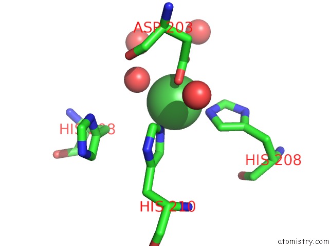
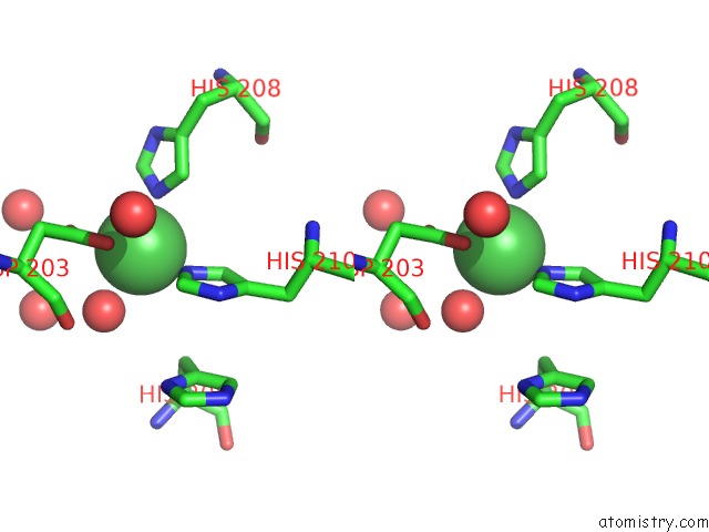
Nickel binding site 1 out of 4 in 5c74
Go back to
Nickel binding site 1 out
of 4 in the Structure of A Novel Protein Arginine Methyltransferase
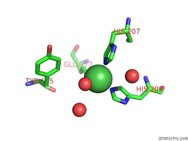
Mono view
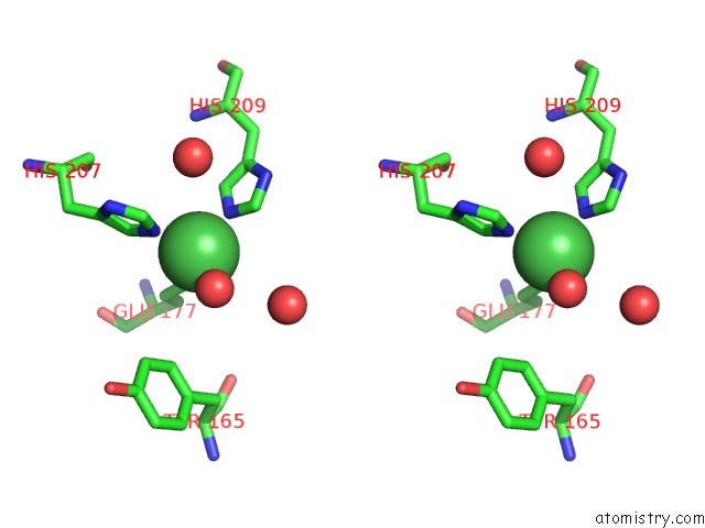
Stereo pair view
Mono view
Stereo pair view
A full contact list of Nickel with other atoms in the Ni binding
site number 1 of Structure of A Novel Protein Arginine Methyltransferase within 5.0Å range:
|
Nickel binding site 2 out of 4 in 5c74
Go back to
Nickel binding site 2 out
of 4 in the Structure of A Novel Protein Arginine Methyltransferase
Mono view
Stereo pair view
Mono view
Stereo pair view
A full contact list of Nickel with other atoms in the Ni binding
site number 2 of Structure of A Novel Protein Arginine Methyltransferase within 5.0Å range:
|
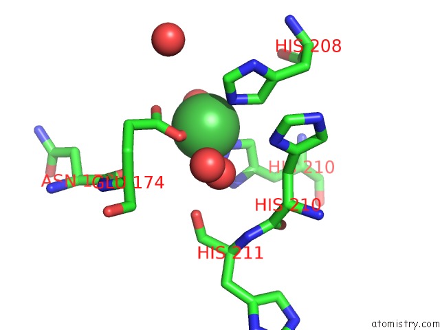
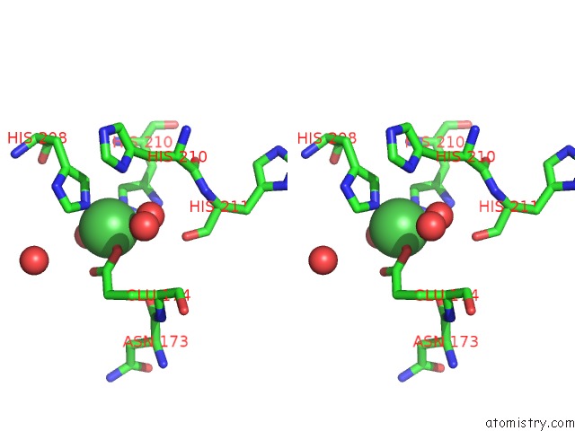
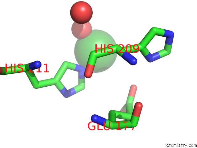
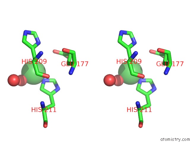
Nickel binding site 3 out of 4 in 5c74
Go back to
Nickel binding site 3 out
of 4 in the Structure of A Novel Protein Arginine Methyltransferase
Mono view
Stereo pair view
Mono view
Stereo pair view
A full contact list of Nickel with other atoms in the Ni binding
site number 3 of Structure of A Novel Protein Arginine Methyltransferase within 5.0Å range:
|
Nickel binding site 4 out of 4 in 5c74
Go back to
Nickel binding site 4 out
of 4 in the Structure of A Novel Protein Arginine Methyltransferase
Mono view
Stereo pair view
Mono view
Stereo pair view
A full contact list of Nickel with other atoms in the Ni binding
site number 4 of Structure of A Novel Protein Arginine Methyltransferase within 5.0Å range:
|
Reference:
F.Lv,
T.Zhang,
Z.Zhou,
S.Gao,
C.Wong,
J.Q.Zhou,
J.Ding.
Structural Basis For SFM1 Functioning As A Protein Arginine Methyltransferase Cell Discov 2015.
ISSN: ESSN 2056-5968
DOI: 10.1038/CELLDISC.2015.37
Page generated: Thu Oct 10 06:16:54 2024
ISSN: ESSN 2056-5968
DOI: 10.1038/CELLDISC.2015.37
Last articles
Zn in 9J0NZn in 9J0O
Zn in 9J0P
Zn in 9FJX
Zn in 9EKB
Zn in 9C0F
Zn in 9CAH
Zn in 9CH0
Zn in 9CH3
Zn in 9CH1