Nickel »
PDB 5bu6-5e6j »
5daw »
Nickel in PDB 5daw: Fe(II)/(Alpha)Ketoglutarate-Dependent Dioxygenase Asqj in Complex with Cyclopeptin
Protein crystallography data
The structure of Fe(II)/(Alpha)Ketoglutarate-Dependent Dioxygenase Asqj in Complex with Cyclopeptin, PDB code: 5daw
was solved by
M.Groll,
A.Braeuer,
with X-Ray Crystallography technique. A brief refinement statistics is given in the table below:
| Resolution Low / High (Å) | 15.00 / 1.60 |
| Space group | C 2 2 21 |
| Cell size a, b, c (Å), α, β, γ (°) | 73.270, 118.600, 67.220, 90.00, 90.00, 90.00 |
| R / Rfree (%) | 18.6 / 19.8 |
Nickel Binding Sites:
The binding sites of Nickel atom in the Fe(II)/(Alpha)Ketoglutarate-Dependent Dioxygenase Asqj in Complex with Cyclopeptin
(pdb code 5daw). This binding sites where shown within
5.0 Angstroms radius around Nickel atom.
In total only one binding site of Nickel was determined in the Fe(II)/(Alpha)Ketoglutarate-Dependent Dioxygenase Asqj in Complex with Cyclopeptin, PDB code: 5daw:
In total only one binding site of Nickel was determined in the Fe(II)/(Alpha)Ketoglutarate-Dependent Dioxygenase Asqj in Complex with Cyclopeptin, PDB code: 5daw:
Nickel binding site 1 out of 1 in 5daw
Go back to
Nickel binding site 1 out
of 1 in the Fe(II)/(Alpha)Ketoglutarate-Dependent Dioxygenase Asqj in Complex with Cyclopeptin
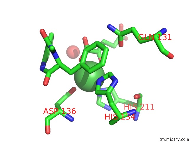
Mono view
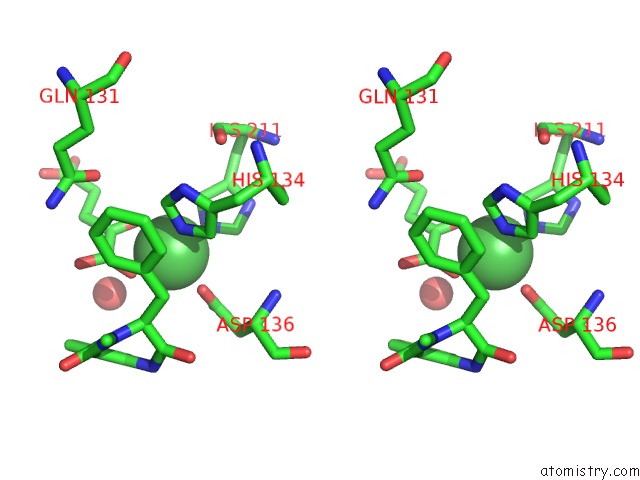
Stereo pair view
Mono view
Stereo pair view
A full contact list of Nickel with other atoms in the Ni binding
site number 1 of Fe(II)/(Alpha)Ketoglutarate-Dependent Dioxygenase Asqj in Complex with Cyclopeptin within 5.0Å range:
|
Reference:
A.Brauer,
P.Beck,
L.Hintermann,
M.Groll.
Structure of the Dioxygenase Asqj: Mechanistic Insights Into A One-Pot Multistep Quinolone Antibiotic Biosynthesis. Angew.Chem.Int.Ed.Engl. V. 55 422 2016.
ISSN: ESSN 1521-3773
PubMed: 26553478
DOI: 10.1002/ANIE.201507835
Page generated: Thu Oct 10 06:19:29 2024
ISSN: ESSN 1521-3773
PubMed: 26553478
DOI: 10.1002/ANIE.201507835
Last articles
Zn in 9J0NZn in 9J0O
Zn in 9J0P
Zn in 9FJX
Zn in 9EKB
Zn in 9C0F
Zn in 9CAH
Zn in 9CH0
Zn in 9CH3
Zn in 9CH1