Nickel »
PDB 6cac-6f5r »
6cu2 »
Nickel in PDB 6cu2: Structure of N-Truncated R2-Type Pyocin Tail Fiber at 2.6 Angstrom Resolution
Protein crystallography data
The structure of Structure of N-Truncated R2-Type Pyocin Tail Fiber at 2.6 Angstrom Resolution, PDB code: 6cu2
was solved by
A.J.Salazar,
M.Sherekar,
J.C.Saccettini,
with X-Ray Crystallography technique. A brief refinement statistics is given in the table below:
| Resolution Low / High (Å) | 39.81 / 2.58 |
| Space group | P 63 2 2 |
| Cell size a, b, c (Å), α, β, γ (°) | 59.865, 59.865, 398.049, 90.00, 90.00, 120.00 |
| R / Rfree (%) | 19.4 / 22.9 |
Other elements in 6cu2:
The structure of Structure of N-Truncated R2-Type Pyocin Tail Fiber at 2.6 Angstrom Resolution also contains other interesting chemical elements:
| Magnesium | (Mg) | 1 atom |
Nickel Binding Sites:
The binding sites of Nickel atom in the Structure of N-Truncated R2-Type Pyocin Tail Fiber at 2.6 Angstrom Resolution
(pdb code 6cu2). This binding sites where shown within
5.0 Angstroms radius around Nickel atom.
In total only one binding site of Nickel was determined in the Structure of N-Truncated R2-Type Pyocin Tail Fiber at 2.6 Angstrom Resolution, PDB code: 6cu2:
In total only one binding site of Nickel was determined in the Structure of N-Truncated R2-Type Pyocin Tail Fiber at 2.6 Angstrom Resolution, PDB code: 6cu2:
Nickel binding site 1 out of 1 in 6cu2
Go back to
Nickel binding site 1 out
of 1 in the Structure of N-Truncated R2-Type Pyocin Tail Fiber at 2.6 Angstrom Resolution
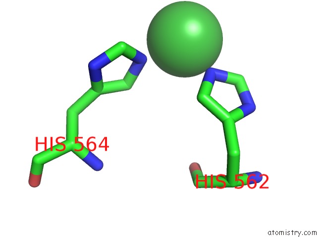
Mono view
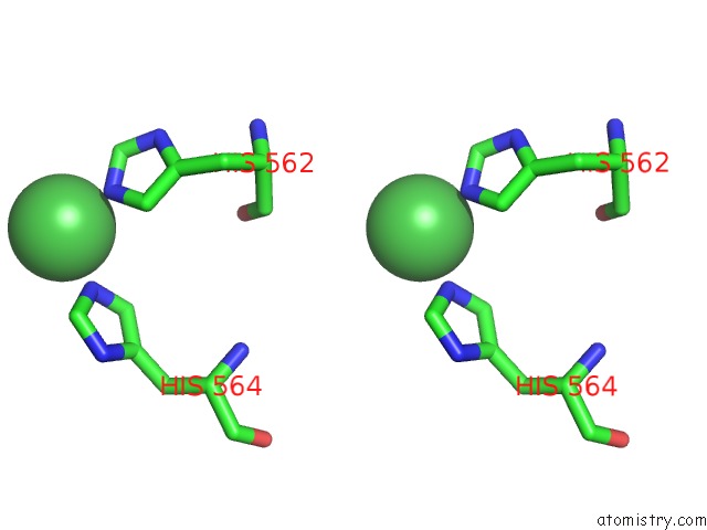
Stereo pair view
Mono view
Stereo pair view
A full contact list of Nickel with other atoms in the Ni binding
site number 1 of Structure of N-Truncated R2-Type Pyocin Tail Fiber at 2.6 Angstrom Resolution within 5.0Å range:
|
Reference:
A.J.Salazar,
M.Sherekar,
J.Tsai,
J.C.Sacchettini.
R Pyocin Tail Fiber Structure Reveals A Receptor-Binding Domain with A Lectin Fold. Plos One V. 14 11432 2019.
ISSN: ESSN 1932-6203
PubMed: 30721244
DOI: 10.1371/JOURNAL.PONE.0211432
Page generated: Thu Oct 10 08:21:13 2024
ISSN: ESSN 1932-6203
PubMed: 30721244
DOI: 10.1371/JOURNAL.PONE.0211432
Last articles
Zn in 9MJ5Zn in 9HNW
Zn in 9G0L
Zn in 9FNE
Zn in 9DZN
Zn in 9E0I
Zn in 9D32
Zn in 9DAK
Zn in 8ZXC
Zn in 8ZUF