Nickel »
PDB 7z5r-8cja »
8ajk »
Nickel in PDB 8ajk: Crystal Structure of A C43S Variant From the Disulfide Reductase Mera From Staphylococcus Aureus
Protein crystallography data
The structure of Crystal Structure of A C43S Variant From the Disulfide Reductase Mera From Staphylococcus Aureus, PDB code: 8ajk
was solved by
P.Weiland,
F.Altegoer,
G.Bange,
with X-Ray Crystallography technique. A brief refinement statistics is given in the table below:
| Resolution Low / High (Å) | 48.68 / 1.60 |
| Space group | P 21 2 21 |
| Cell size a, b, c (Å), α, β, γ (°) | 61.25, 88.79, 186.84, 90, 90, 90 |
| R / Rfree (%) | 17.8 / 20.3 |
Nickel Binding Sites:
The binding sites of Nickel atom in the Crystal Structure of A C43S Variant From the Disulfide Reductase Mera From Staphylococcus Aureus
(pdb code 8ajk). This binding sites where shown within
5.0 Angstroms radius around Nickel atom.
In total 2 binding sites of Nickel where determined in the Crystal Structure of A C43S Variant From the Disulfide Reductase Mera From Staphylococcus Aureus, PDB code: 8ajk:
Jump to Nickel binding site number: 1; 2;
In total 2 binding sites of Nickel where determined in the Crystal Structure of A C43S Variant From the Disulfide Reductase Mera From Staphylococcus Aureus, PDB code: 8ajk:
Jump to Nickel binding site number: 1; 2;
Nickel binding site 1 out of 2 in 8ajk
Go back to
Nickel binding site 1 out
of 2 in the Crystal Structure of A C43S Variant From the Disulfide Reductase Mera From Staphylococcus Aureus

Mono view
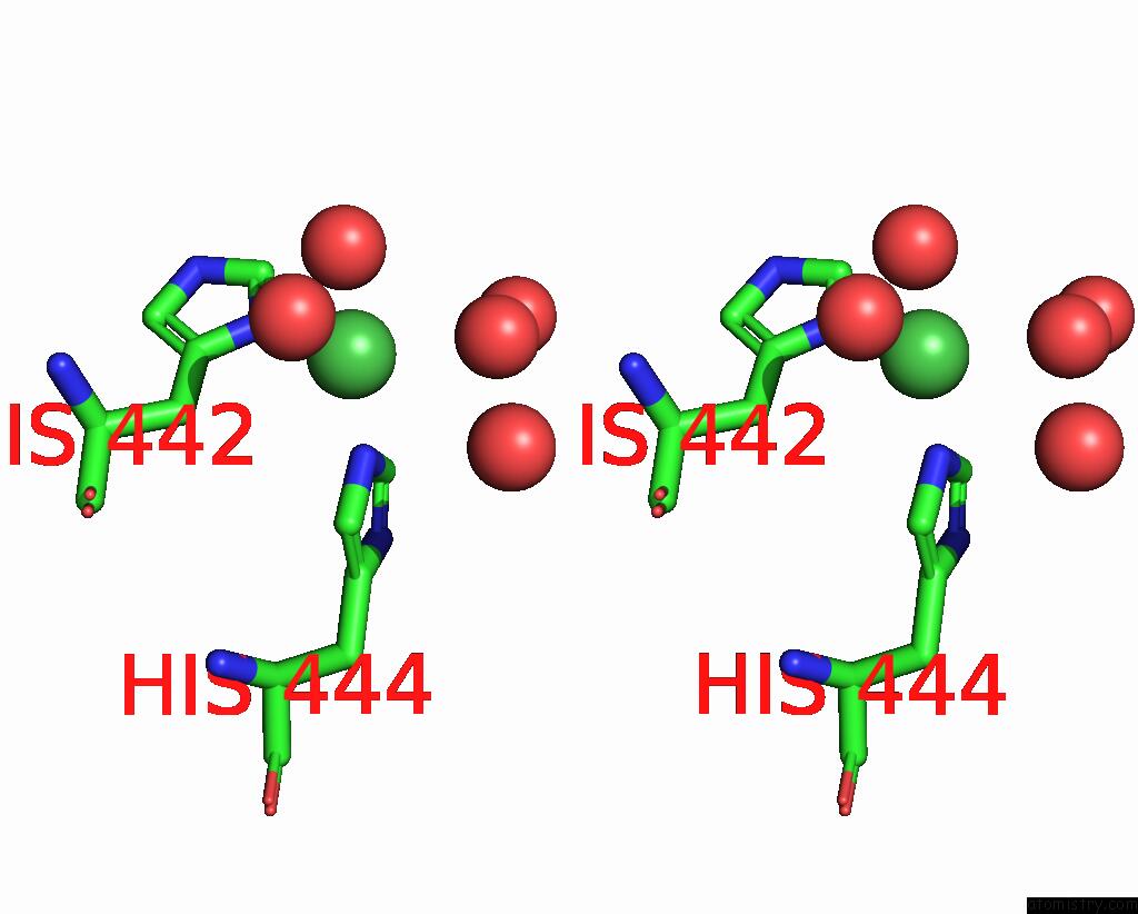
Stereo pair view
Mono view
Stereo pair view
A full contact list of Nickel with other atoms in the Ni binding
site number 1 of Crystal Structure of A C43S Variant From the Disulfide Reductase Mera From Staphylococcus Aureus within 5.0Å range:
|
Nickel binding site 2 out of 2 in 8ajk
Go back to
Nickel binding site 2 out
of 2 in the Crystal Structure of A C43S Variant From the Disulfide Reductase Mera From Staphylococcus Aureus
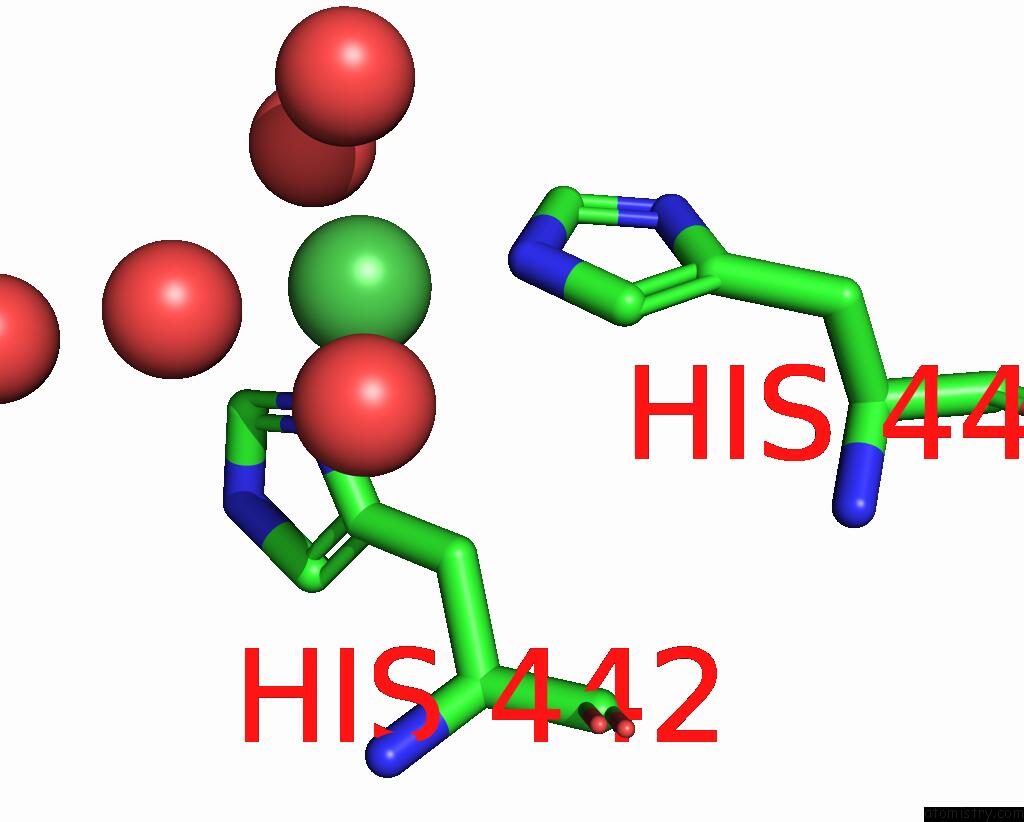
Mono view
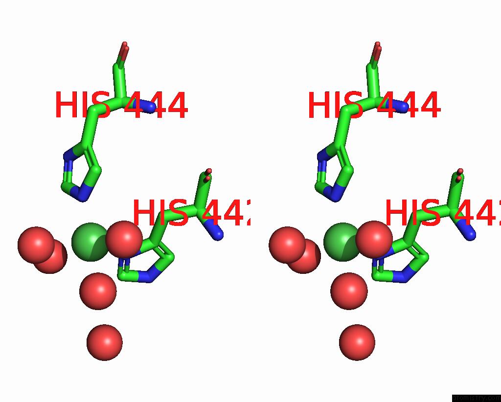
Stereo pair view
Mono view
Stereo pair view
A full contact list of Nickel with other atoms in the Ni binding
site number 2 of Crystal Structure of A C43S Variant From the Disulfide Reductase Mera From Staphylococcus Aureus within 5.0Å range:
|
Reference:
H.L.Shearer,
V.Van Loi,
P.Weiland,
G.Bange,
F.Altegoer,
M.B.Hampton,
H.Antelmann,
N.Dickerhof.
Mera Functions As A Hypothiocyanous Acid Reductase and Defense Mechanism in Staphylococcus Aureus. Mol.Microbiol. 2023.
ISSN: ESSN 1365-2958
PubMed: 36779383
DOI: 10.1111/MMI.15035
Page generated: Thu Oct 10 09:34:54 2024
ISSN: ESSN 1365-2958
PubMed: 36779383
DOI: 10.1111/MMI.15035
Last articles
Zn in 9J0NZn in 9J0O
Zn in 9J0P
Zn in 9FJX
Zn in 9EKB
Zn in 9C0F
Zn in 9CAH
Zn in 9CH0
Zn in 9CH3
Zn in 9CH1