Nickel »
PDB 7z5r-8cja »
8amq »
Nickel in PDB 8amq: Crystal Structure of the Complex CYP143-Fdxe From M. Tuberculosis
Protein crystallography data
The structure of Crystal Structure of the Complex CYP143-Fdxe From M. Tuberculosis, PDB code: 8amq
was solved by
S.Bukhdruker,
T.Varaksa,
S.Smolskaya,
E.Marin,
I.Kapranov,
K.Kovalev,
A.Gilep,
N.Strushkevich,
V.Borshchevskiy,
with X-Ray Crystallography technique. A brief refinement statistics is given in the table below:
| Resolution Low / High (Å) | 27.15 / 1.60 |
| Space group | P 1 |
| Cell size a, b, c (Å), α, β, γ (°) | 53.2, 54.354, 69.037, 67.71, 77.21, 61.63 |
| R / Rfree (%) | 17.7 / 20.7 |
Other elements in 8amq:
The structure of Crystal Structure of the Complex CYP143-Fdxe From M. Tuberculosis also contains other interesting chemical elements:
| Iron | (Fe) | 4 atoms |
Nickel Binding Sites:
The binding sites of Nickel atom in the Crystal Structure of the Complex CYP143-Fdxe From M. Tuberculosis
(pdb code 8amq). This binding sites where shown within
5.0 Angstroms radius around Nickel atom.
In total 6 binding sites of Nickel where determined in the Crystal Structure of the Complex CYP143-Fdxe From M. Tuberculosis, PDB code: 8amq:
Jump to Nickel binding site number: 1; 2; 3; 4; 5; 6;
In total 6 binding sites of Nickel where determined in the Crystal Structure of the Complex CYP143-Fdxe From M. Tuberculosis, PDB code: 8amq:
Jump to Nickel binding site number: 1; 2; 3; 4; 5; 6;
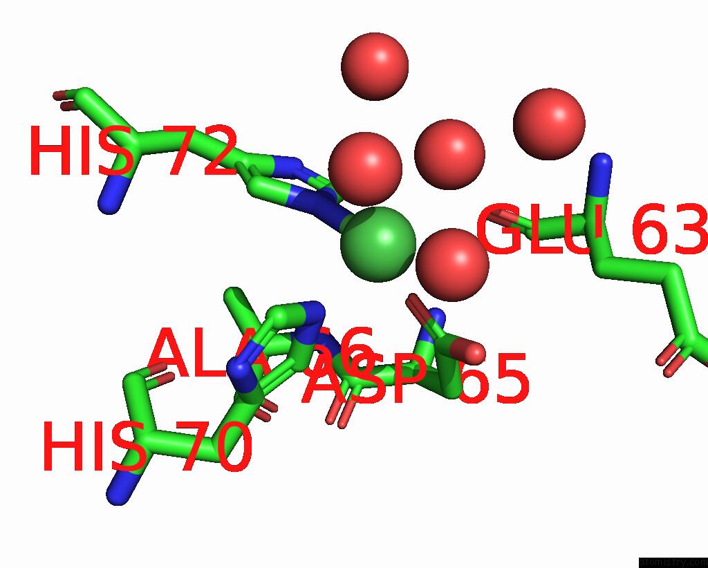
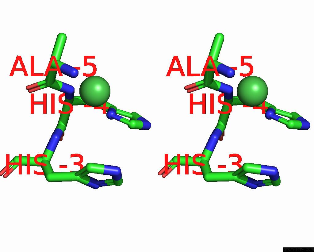
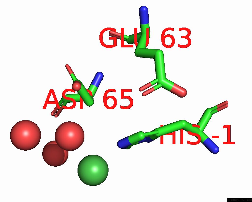
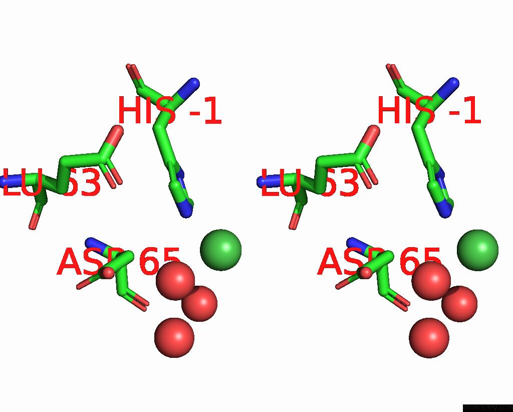
Nickel binding site 1 out of 6 in 8amq
Go back to
Nickel binding site 1 out
of 6 in the Crystal Structure of the Complex CYP143-Fdxe From M. Tuberculosis
Mono view
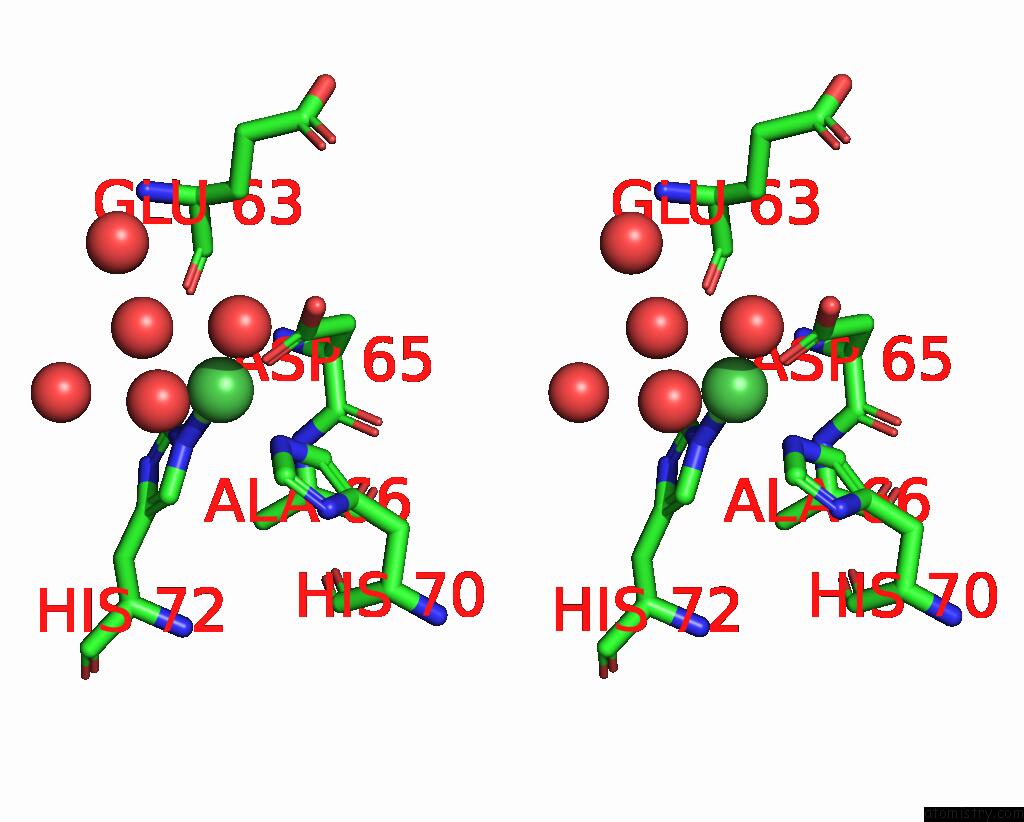
Stereo pair view
Mono view
Stereo pair view
A full contact list of Nickel with other atoms in the Ni binding
site number 1 of Crystal Structure of the Complex CYP143-Fdxe From M. Tuberculosis within 5.0Å range:
|
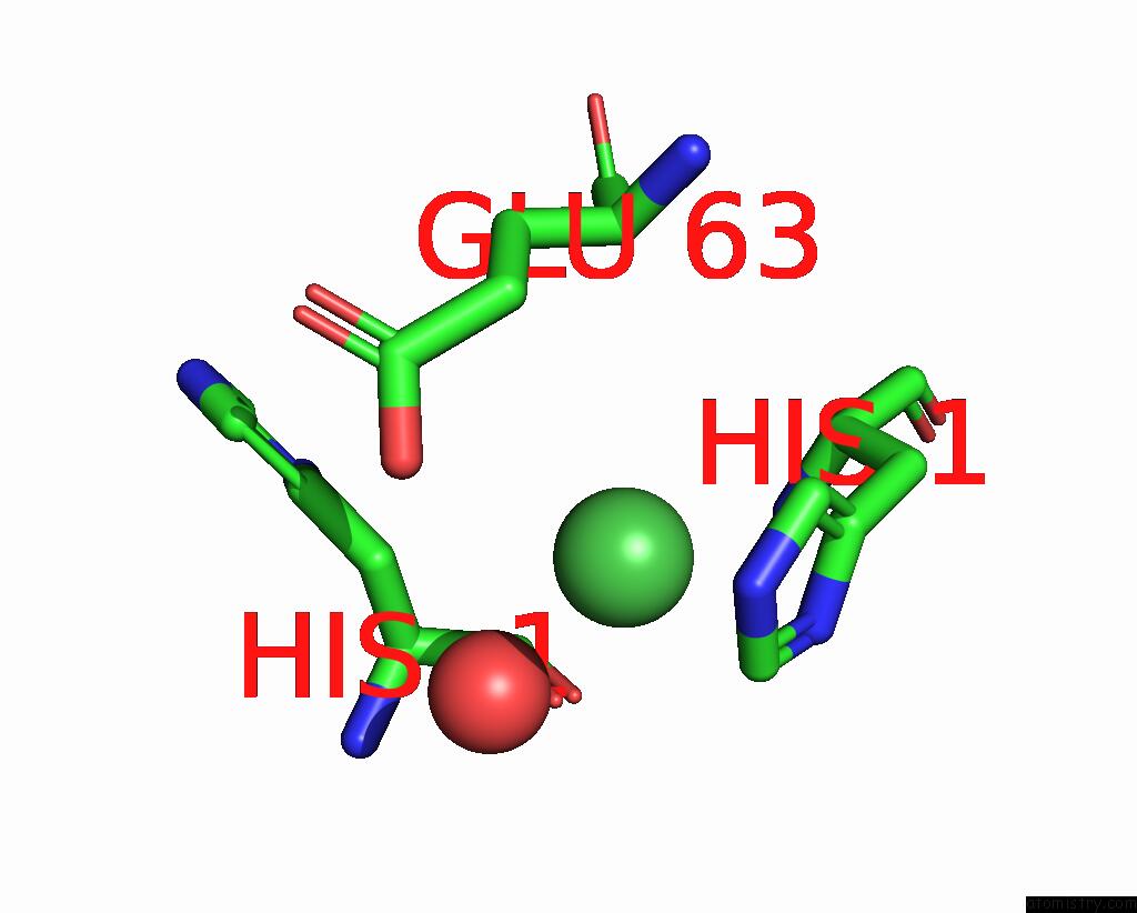
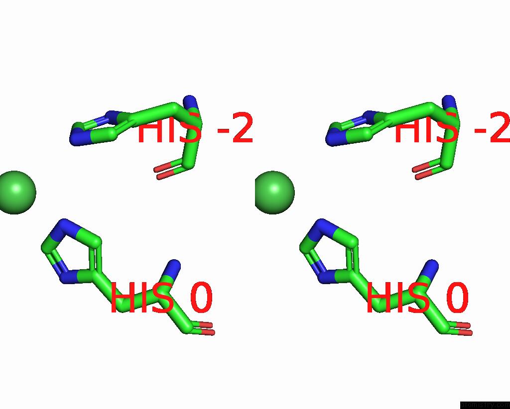
Nickel binding site 2 out of 6 in 8amq
Go back to
Nickel binding site 2 out
of 6 in the Crystal Structure of the Complex CYP143-Fdxe From M. Tuberculosis
Mono view

Stereo pair view
Mono view
Stereo pair view
A full contact list of Nickel with other atoms in the Ni binding
site number 2 of Crystal Structure of the Complex CYP143-Fdxe From M. Tuberculosis within 5.0Å range:
|
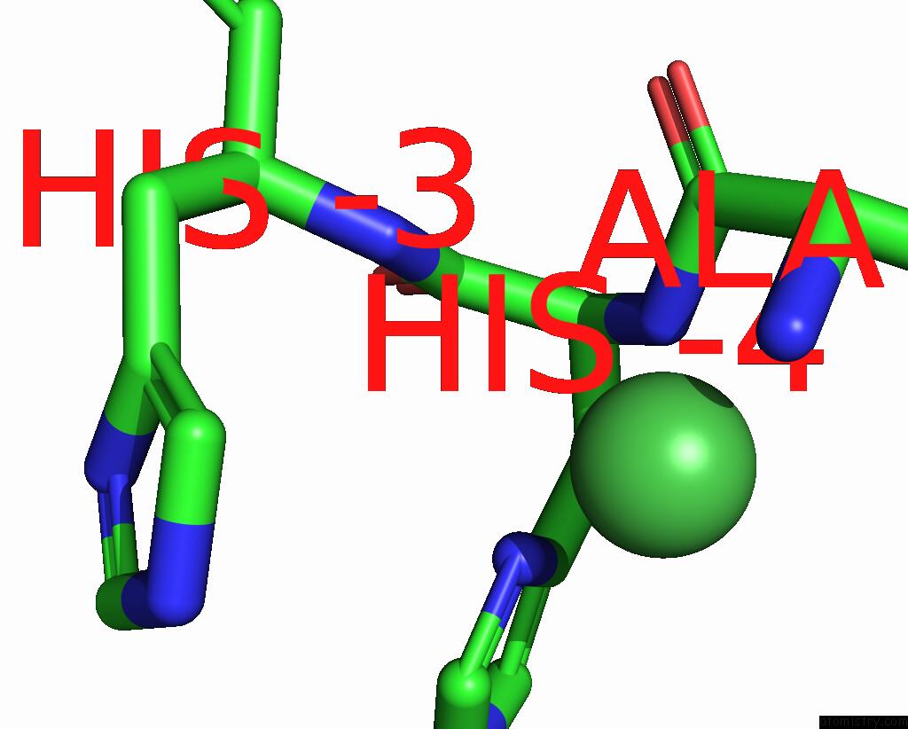
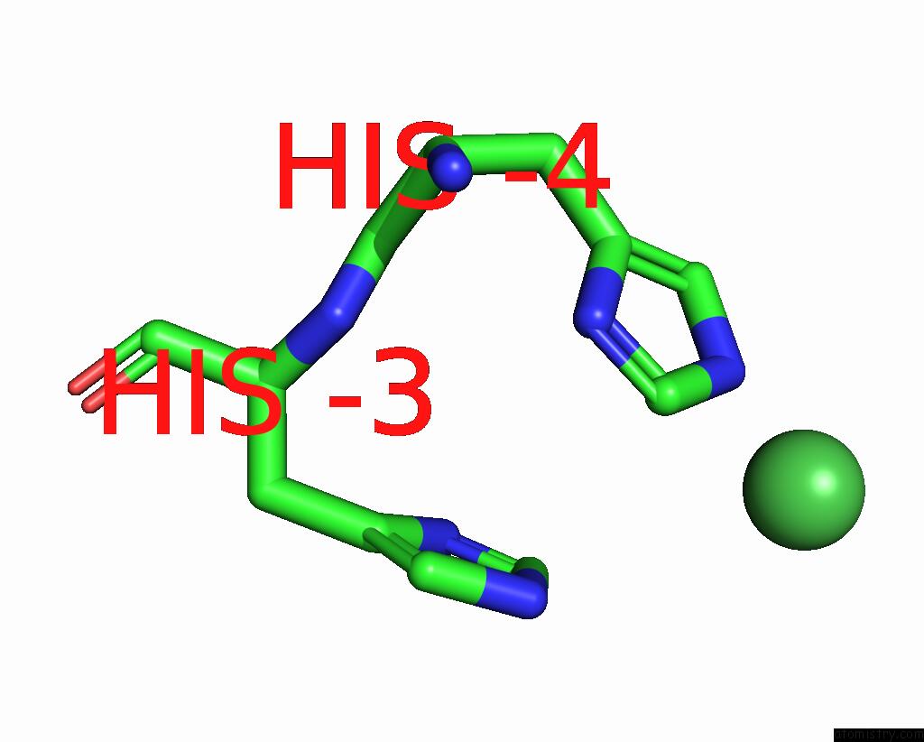
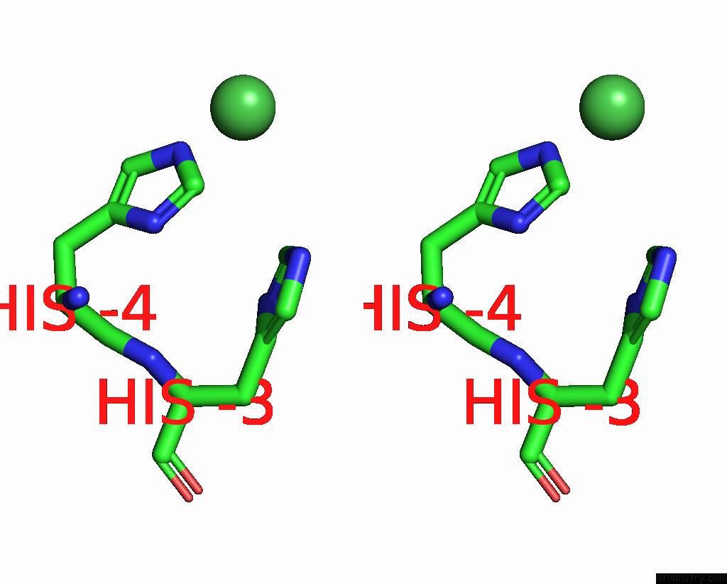
Nickel binding site 3 out of 6 in 8amq
Go back to
Nickel binding site 3 out
of 6 in the Crystal Structure of the Complex CYP143-Fdxe From M. Tuberculosis
Mono view
Stereo pair view
Mono view
Stereo pair view
A full contact list of Nickel with other atoms in the Ni binding
site number 3 of Crystal Structure of the Complex CYP143-Fdxe From M. Tuberculosis within 5.0Å range:
|
Nickel binding site 4 out of 6 in 8amq
Go back to
Nickel binding site 4 out
of 6 in the Crystal Structure of the Complex CYP143-Fdxe From M. Tuberculosis

Mono view
Stereo pair view
Mono view
Stereo pair view
A full contact list of Nickel with other atoms in the Ni binding
site number 4 of Crystal Structure of the Complex CYP143-Fdxe From M. Tuberculosis within 5.0Å range:
|
Nickel binding site 5 out of 6 in 8amq
Go back to
Nickel binding site 5 out
of 6 in the Crystal Structure of the Complex CYP143-Fdxe From M. Tuberculosis
Mono view
Stereo pair view
Mono view
Stereo pair view
A full contact list of Nickel with other atoms in the Ni binding
site number 5 of Crystal Structure of the Complex CYP143-Fdxe From M. Tuberculosis within 5.0Å range:
|
Nickel binding site 6 out of 6 in 8amq
Go back to
Nickel binding site 6 out
of 6 in the Crystal Structure of the Complex CYP143-Fdxe From M. Tuberculosis
Mono view
Stereo pair view
Mono view
Stereo pair view
A full contact list of Nickel with other atoms in the Ni binding
site number 6 of Crystal Structure of the Complex CYP143-Fdxe From M. Tuberculosis within 5.0Å range:
|
Reference:
A.Gilep,
T.Varaksa,
S.Bukhdruker,
A.Kavaleuski,
Y.Ryzhykau,
S.Smolskaya,
T.Sushko,
K.Tsumoto,
I.Grabovec,
I.Kapranov,
I.Okhrimenko,
E.Marin,
M.Shevtsov,
A.Mishin,
K.Kovalev,
A.Kuklin,
V.Gordeliy,
L.Kaluzhskiy,
O.Gnedenko,
E.Yablokov,
A.Ivanov,
V.Borshchevskiy,
N.Strushkevich.
Structural Insights Into 3FE-4S Ferredoxins Diversity in M. Tuberculosis Highlighted By A First Redox Complex with P450. Front Mol Biosci V. 9 00032 2022.
ISSN: ESSN 2296-889X
PubMed: 36699703
DOI: 10.3389/FMOLB.2022.1100032
Page generated: Thu Oct 10 09:34:59 2024
ISSN: ESSN 2296-889X
PubMed: 36699703
DOI: 10.3389/FMOLB.2022.1100032
Last articles
Zn in 9MJ5Zn in 9HNW
Zn in 9G0L
Zn in 9FNE
Zn in 9DZN
Zn in 9E0I
Zn in 9D32
Zn in 9DAK
Zn in 8ZXC
Zn in 8ZUF