Nickel »
PDB 7z5r-8cja »
8bj9 »
Nickel in PDB 8bj9: X-Ray Structure of the Ceue Homologue From Parageobacillus Thermoglucosidasius - 5LICAM Complex.
Protein crystallography data
The structure of X-Ray Structure of the Ceue Homologue From Parageobacillus Thermoglucosidasius - 5LICAM Complex., PDB code: 8bj9
was solved by
E.V.Blagova,
M.Bennett,
R.Booth,
E.J.Dodson,
A.-K.Duhme-Klair,
K.S.Wilson,
with X-Ray Crystallography technique. A brief refinement statistics is given in the table below:
| Resolution Low / High (Å) | 36.83 / 2.07 |
| Space group | C 2 2 21 |
| Cell size a, b, c (Å), α, β, γ (°) | 36.063, 117.392, 141.712, 90, 90, 90 |
| R / Rfree (%) | 21 / 26.6 |
Other elements in 8bj9:
The structure of X-Ray Structure of the Ceue Homologue From Parageobacillus Thermoglucosidasius - 5LICAM Complex. also contains other interesting chemical elements:
| Iron | (Fe) | 1 atom |
Nickel Binding Sites:
The binding sites of Nickel atom in the X-Ray Structure of the Ceue Homologue From Parageobacillus Thermoglucosidasius - 5LICAM Complex.
(pdb code 8bj9). This binding sites where shown within
5.0 Angstroms radius around Nickel atom.
In total 4 binding sites of Nickel where determined in the X-Ray Structure of the Ceue Homologue From Parageobacillus Thermoglucosidasius - 5LICAM Complex., PDB code: 8bj9:
Jump to Nickel binding site number: 1; 2; 3; 4;
In total 4 binding sites of Nickel where determined in the X-Ray Structure of the Ceue Homologue From Parageobacillus Thermoglucosidasius - 5LICAM Complex., PDB code: 8bj9:
Jump to Nickel binding site number: 1; 2; 3; 4;
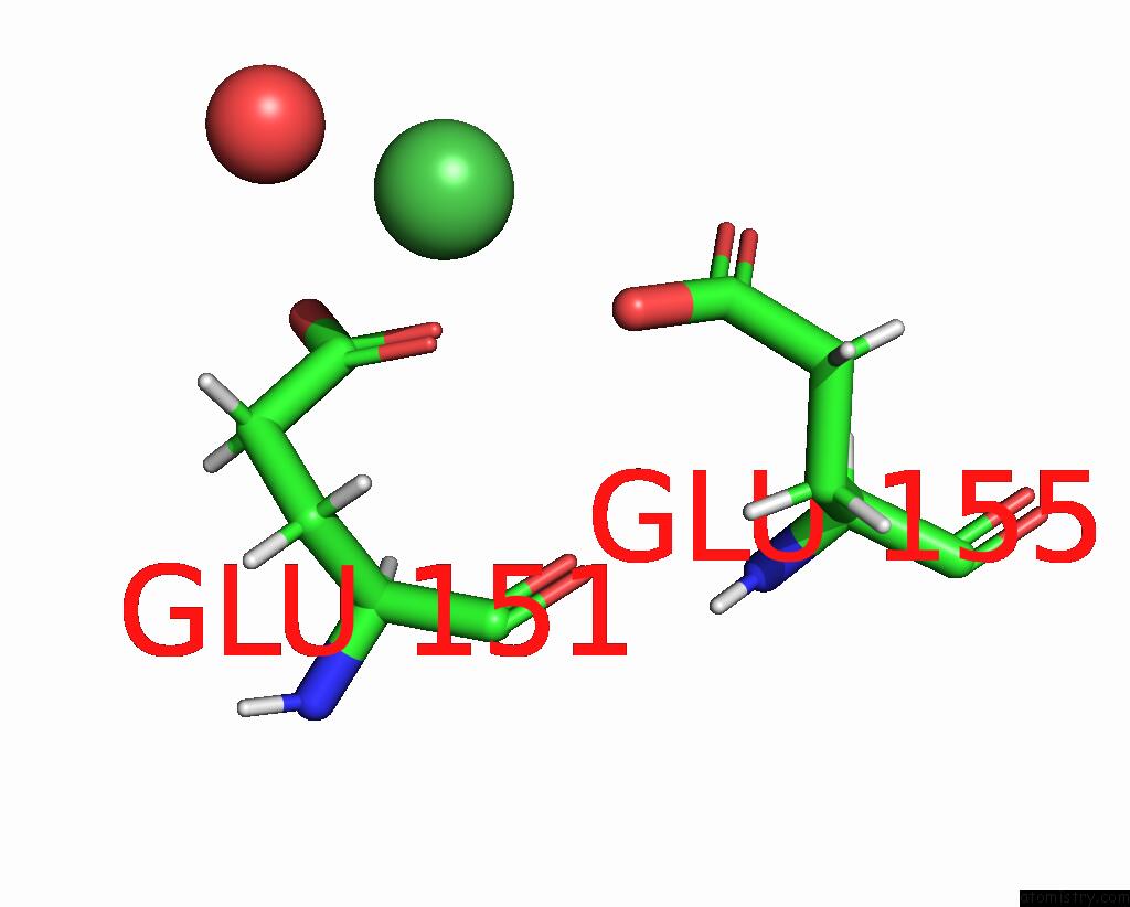
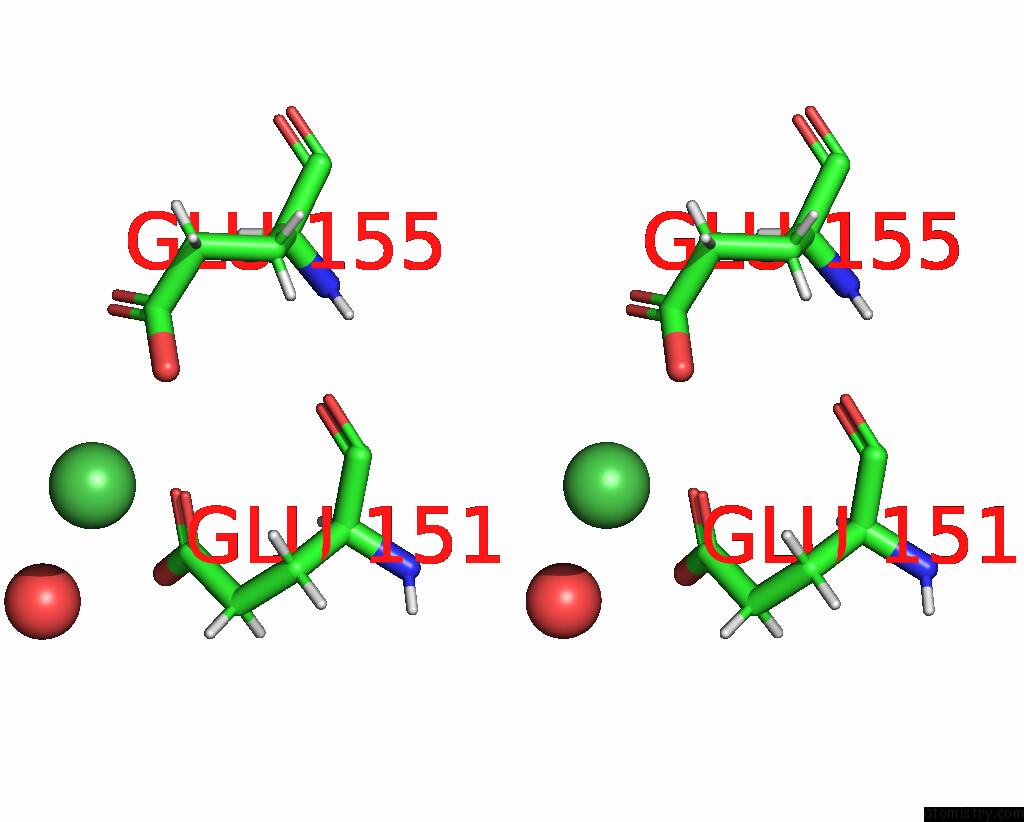
Nickel binding site 1 out of 4 in 8bj9
Go back to
Nickel binding site 1 out
of 4 in the X-Ray Structure of the Ceue Homologue From Parageobacillus Thermoglucosidasius - 5LICAM Complex.
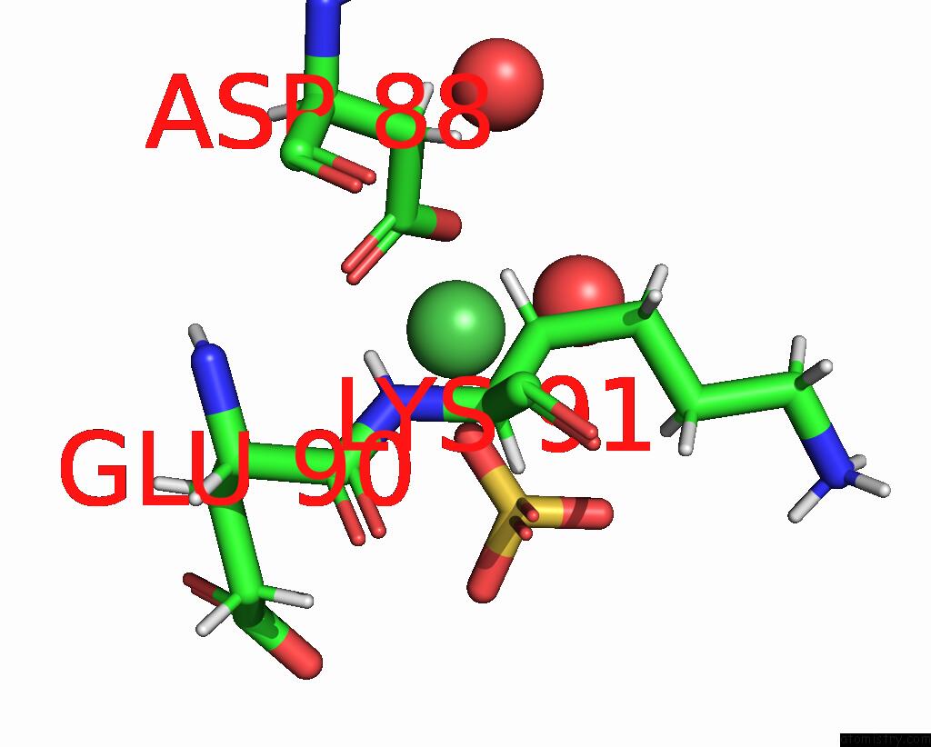
Mono view
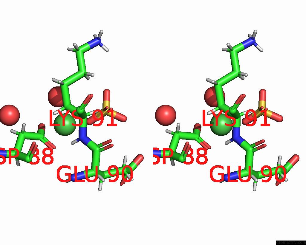
Stereo pair view
Mono view
Stereo pair view
A full contact list of Nickel with other atoms in the Ni binding
site number 1 of X-Ray Structure of the Ceue Homologue From Parageobacillus Thermoglucosidasius - 5LICAM Complex. within 5.0Å range:
|
Nickel binding site 2 out of 4 in 8bj9
Go back to
Nickel binding site 2 out
of 4 in the X-Ray Structure of the Ceue Homologue From Parageobacillus Thermoglucosidasius - 5LICAM Complex.
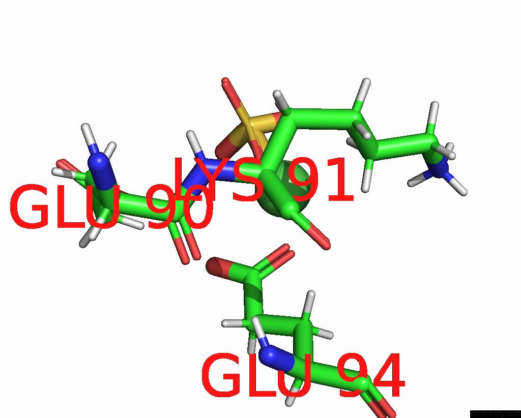
Mono view
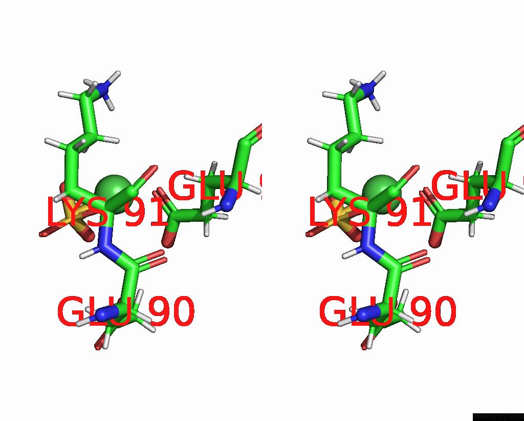
Stereo pair view
Mono view
Stereo pair view
A full contact list of Nickel with other atoms in the Ni binding
site number 2 of X-Ray Structure of the Ceue Homologue From Parageobacillus Thermoglucosidasius - 5LICAM Complex. within 5.0Å range:
|
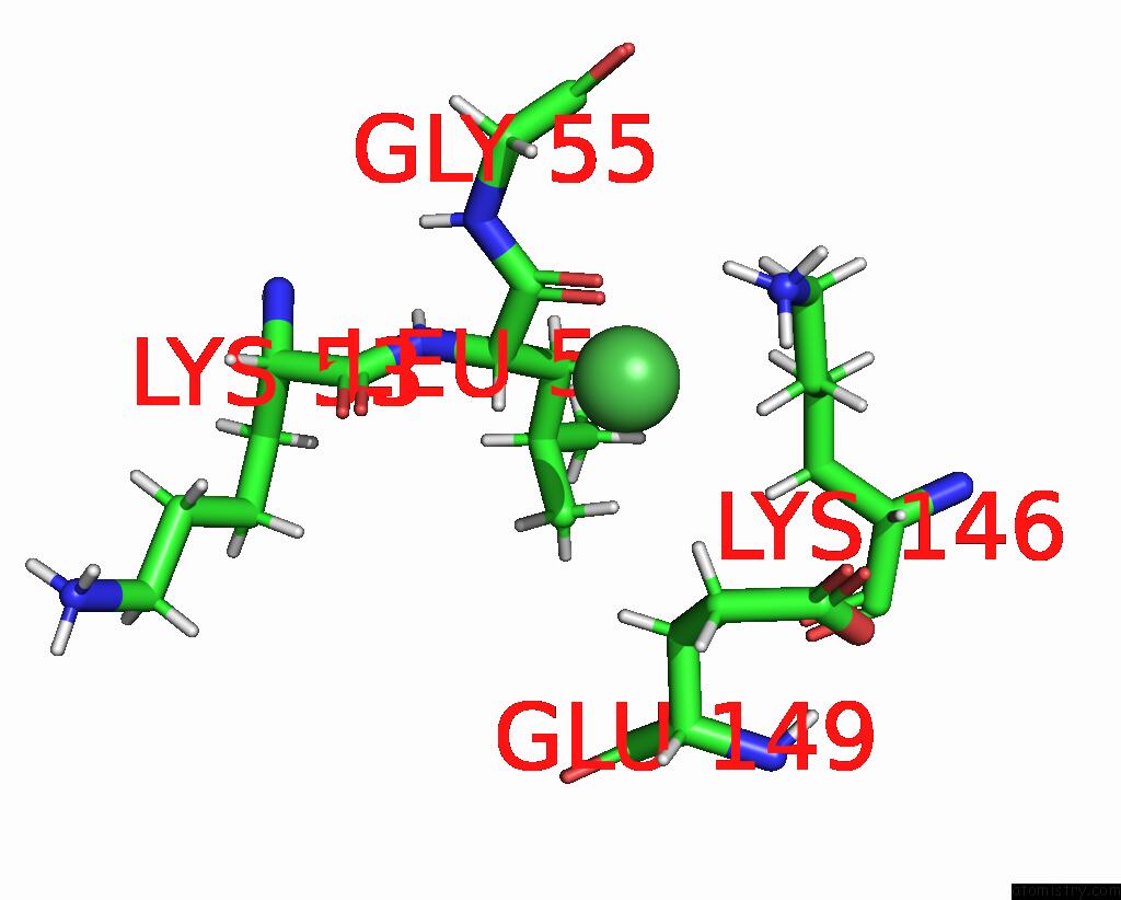
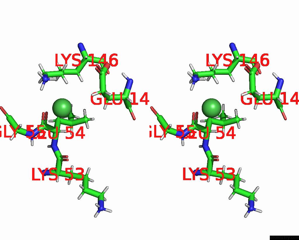
Nickel binding site 3 out of 4 in 8bj9
Go back to
Nickel binding site 3 out
of 4 in the X-Ray Structure of the Ceue Homologue From Parageobacillus Thermoglucosidasius - 5LICAM Complex.
Mono view
Stereo pair view
Mono view
Stereo pair view
A full contact list of Nickel with other atoms in the Ni binding
site number 3 of X-Ray Structure of the Ceue Homologue From Parageobacillus Thermoglucosidasius - 5LICAM Complex. within 5.0Å range:
|
Nickel binding site 4 out of 4 in 8bj9
Go back to
Nickel binding site 4 out
of 4 in the X-Ray Structure of the Ceue Homologue From Parageobacillus Thermoglucosidasius - 5LICAM Complex.
Mono view
Stereo pair view
Mono view
Stereo pair view
A full contact list of Nickel with other atoms in the Ni binding
site number 4 of X-Ray Structure of the Ceue Homologue From Parageobacillus Thermoglucosidasius - 5LICAM Complex. within 5.0Å range:
|
Reference:
E.V.Blagova,
A.Miller,
M.Bennett,
R.Booth,
E.J.Dodson,
A.-K.Duhme-Klair,
K.S.Wilson.
Thermostable Homologues of the Periplasmic Siderophore-Binding Protein Ceue From and Acta Crystallogr.,Sect.D 2023.
ISSN: ESSN 1399-0047
Page generated: Thu Oct 10 09:36:38 2024
ISSN: ESSN 1399-0047
Last articles
Zn in 9J0NZn in 9J0O
Zn in 9J0P
Zn in 9FJX
Zn in 9EKB
Zn in 9C0F
Zn in 9CAH
Zn in 9CH0
Zn in 9CH3
Zn in 9CH1