Nickel »
PDB 8oi7-8xc2 »
8sp2 »
Nickel in PDB 8sp2: Crystal Structure of Metformin Hydrolase (Mfmab) From Pseudomonas Mendocina Sp. Met-2 Apo Form
Protein crystallography data
The structure of Crystal Structure of Metformin Hydrolase (Mfmab) From Pseudomonas Mendocina Sp. Met-2 Apo Form, PDB code: 8sp2
was solved by
L.J.Tassoulas,
J.A.Rankin,
M.H.Elias,
L.P.Wackett,
with X-Ray Crystallography technique. A brief refinement statistics is given in the table below:
| Resolution Low / High (Å) | 74.61 / 2.20 |
| Space group | P 1 |
| Cell size a, b, c (Å), α, β, γ (°) | 103.53, 107.88, 114.51, 93.43, 97.63, 98.11 |
| R / Rfree (%) | 21.3 / 25.7 |
Nickel Binding Sites:
The binding sites of Nickel atom in the Crystal Structure of Metformin Hydrolase (Mfmab) From Pseudomonas Mendocina Sp. Met-2 Apo Form
(pdb code 8sp2). This binding sites where shown within
5.0 Angstroms radius around Nickel atom.
In total 8 binding sites of Nickel where determined in the Crystal Structure of Metformin Hydrolase (Mfmab) From Pseudomonas Mendocina Sp. Met-2 Apo Form, PDB code: 8sp2:
Jump to Nickel binding site number: 1; 2; 3; 4; 5; 6; 7; 8;
In total 8 binding sites of Nickel where determined in the Crystal Structure of Metformin Hydrolase (Mfmab) From Pseudomonas Mendocina Sp. Met-2 Apo Form, PDB code: 8sp2:
Jump to Nickel binding site number: 1; 2; 3; 4; 5; 6; 7; 8;
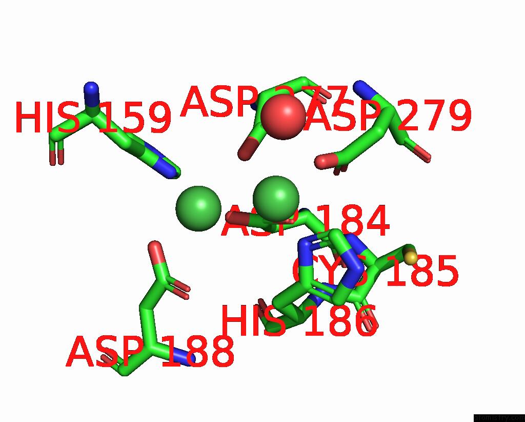
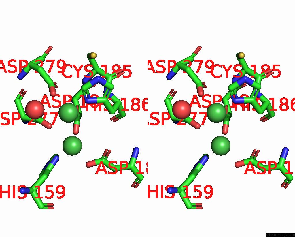
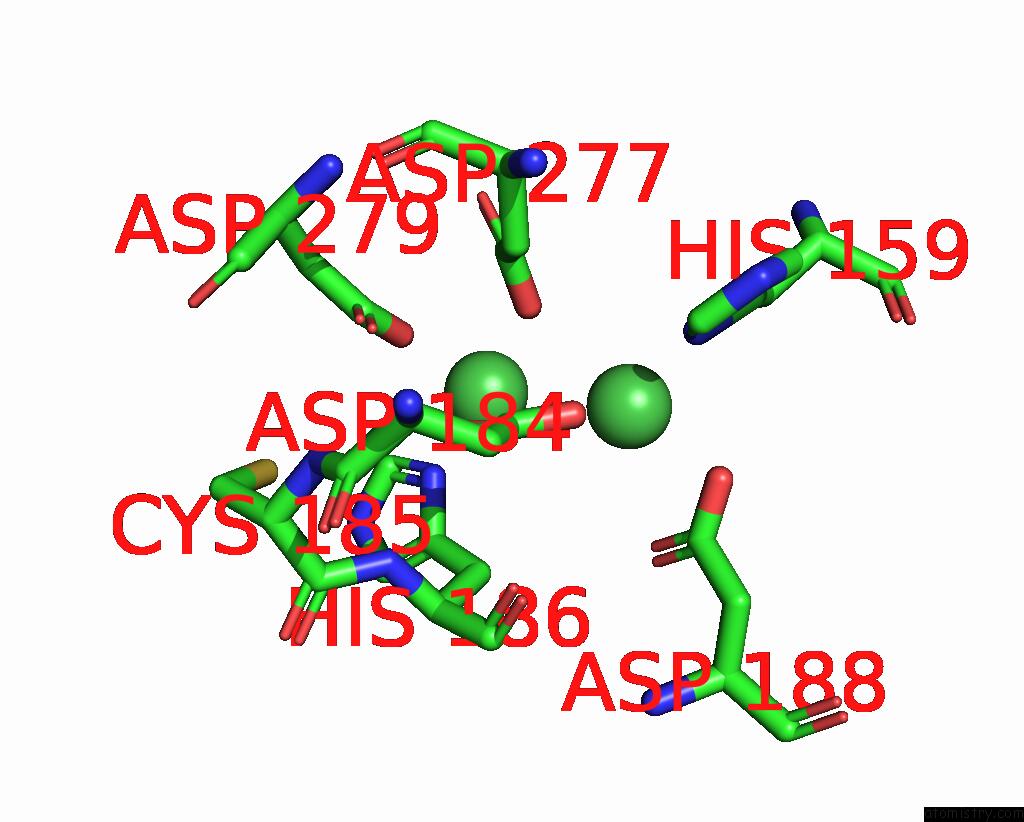
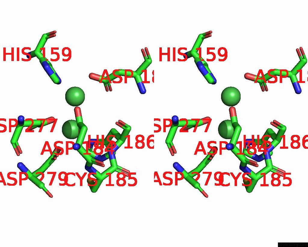
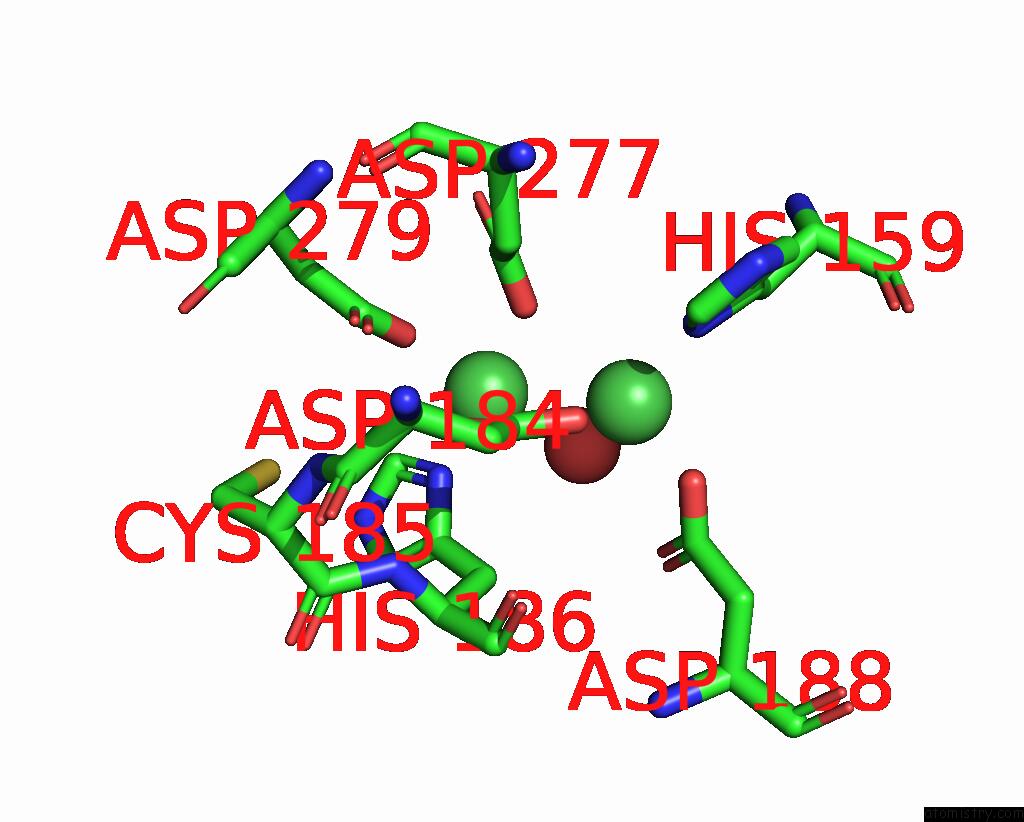
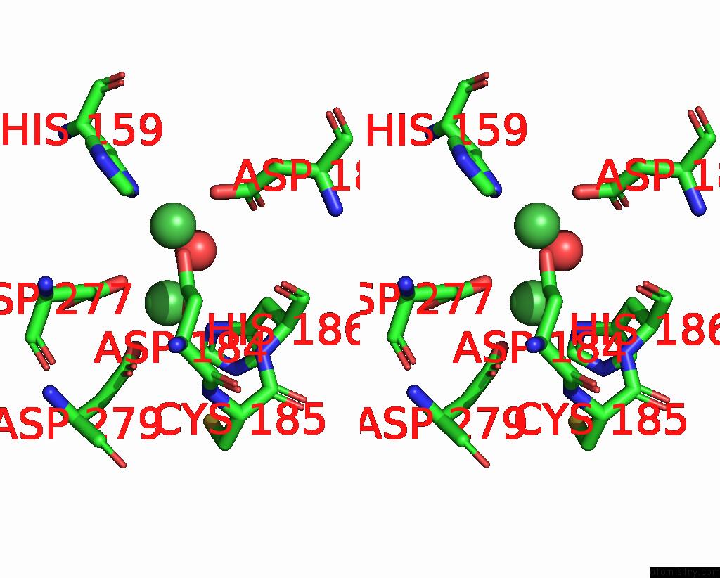
Nickel binding site 1 out of 8 in 8sp2
Go back to
Nickel binding site 1 out
of 8 in the Crystal Structure of Metformin Hydrolase (Mfmab) From Pseudomonas Mendocina Sp. Met-2 Apo Form
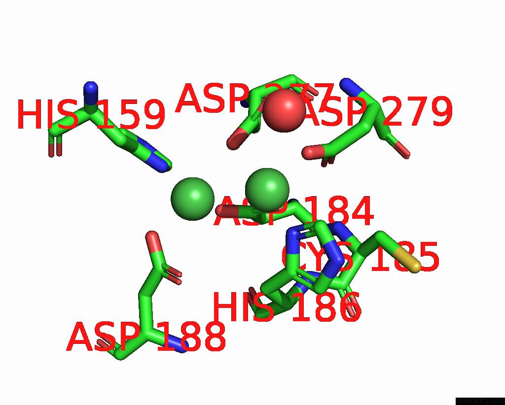
Mono view
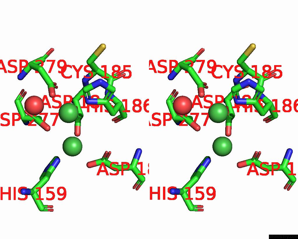
Stereo pair view
Mono view
Stereo pair view
A full contact list of Nickel with other atoms in the Ni binding
site number 1 of Crystal Structure of Metformin Hydrolase (Mfmab) From Pseudomonas Mendocina Sp. Met-2 Apo Form within 5.0Å range:
|
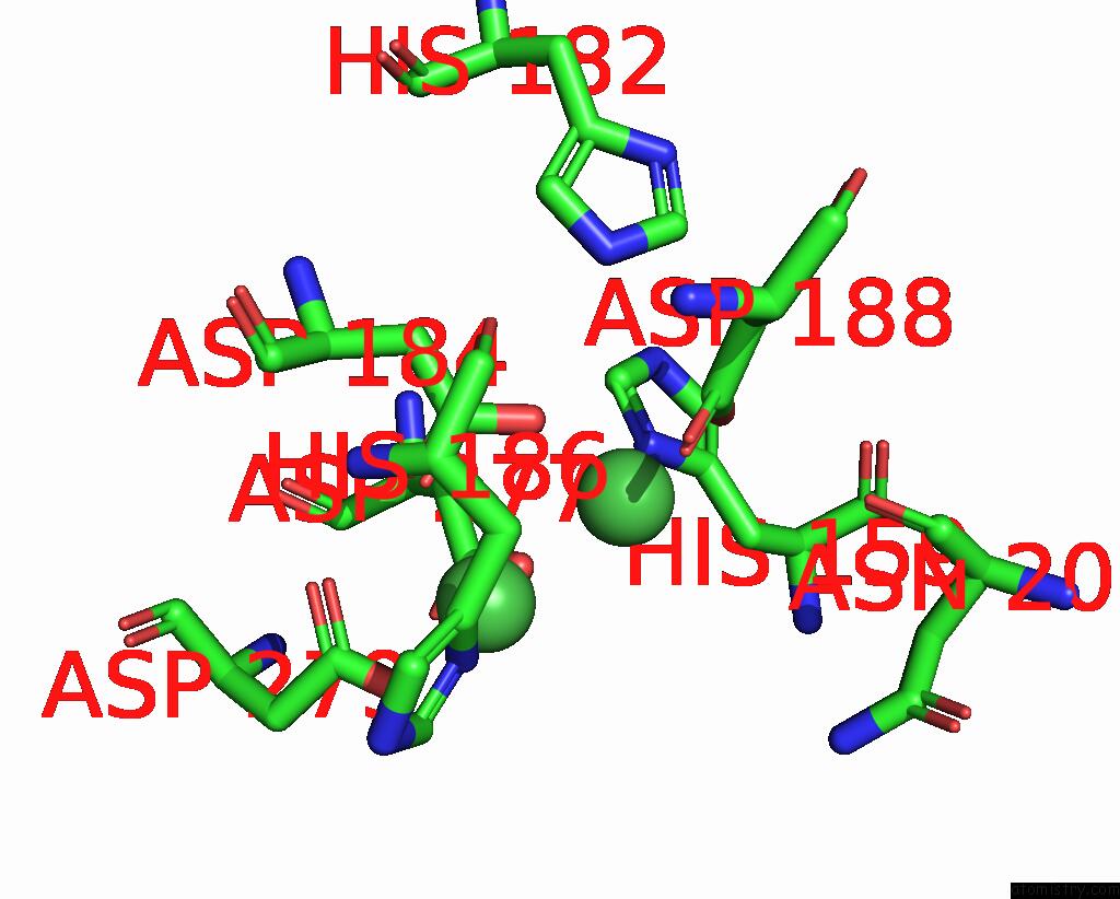
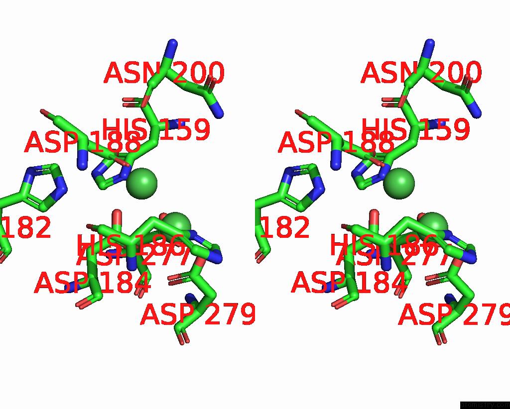
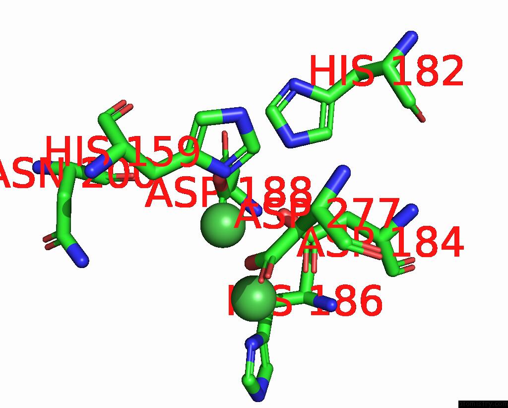
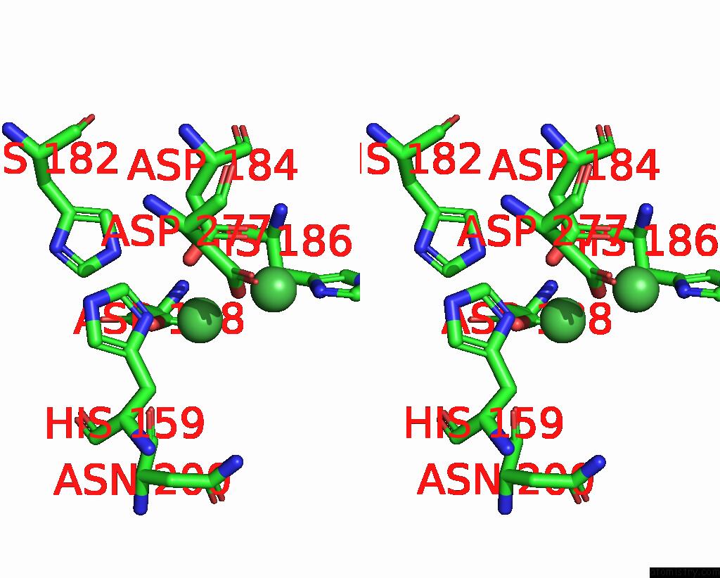
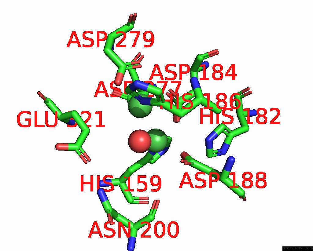
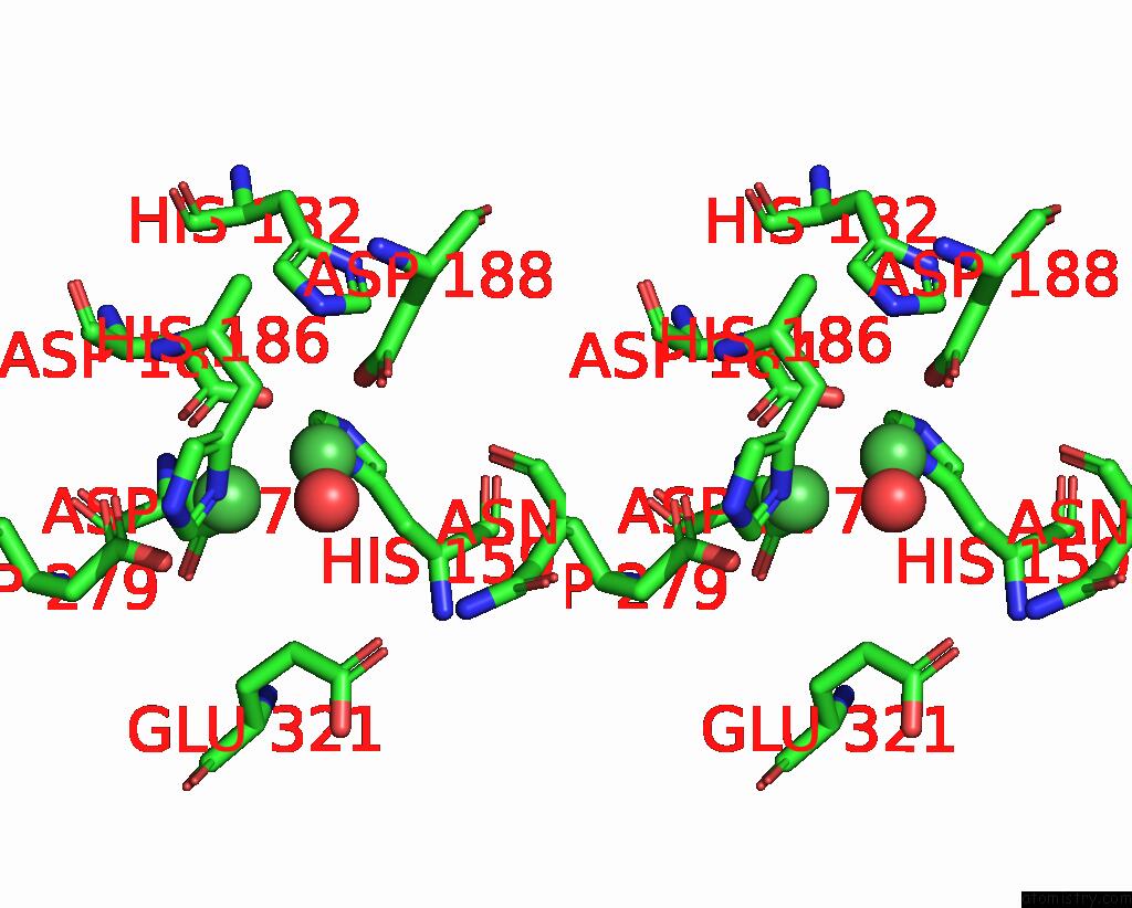
Nickel binding site 2 out of 8 in 8sp2
Go back to
Nickel binding site 2 out
of 8 in the Crystal Structure of Metformin Hydrolase (Mfmab) From Pseudomonas Mendocina Sp. Met-2 Apo Form
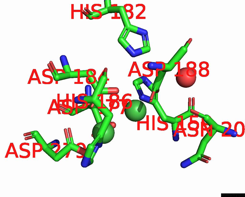
Mono view
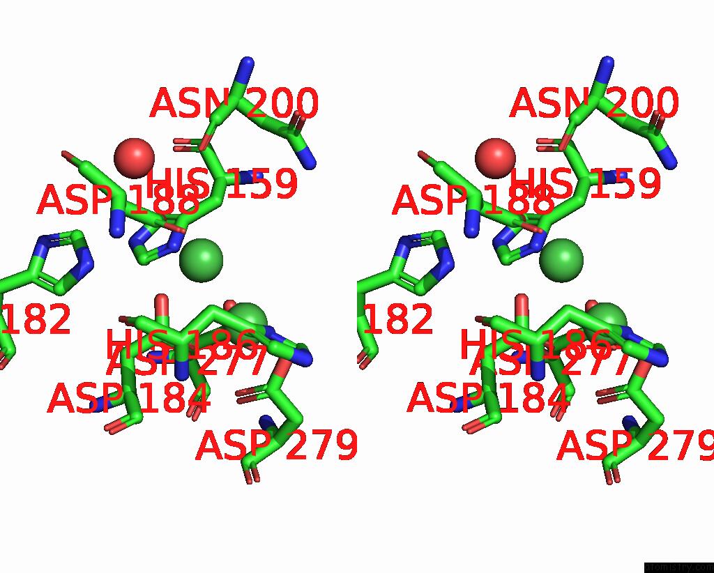
Stereo pair view
Mono view
Stereo pair view
A full contact list of Nickel with other atoms in the Ni binding
site number 2 of Crystal Structure of Metformin Hydrolase (Mfmab) From Pseudomonas Mendocina Sp. Met-2 Apo Form within 5.0Å range:
|
Nickel binding site 3 out of 8 in 8sp2
Go back to
Nickel binding site 3 out
of 8 in the Crystal Structure of Metformin Hydrolase (Mfmab) From Pseudomonas Mendocina Sp. Met-2 Apo Form
Mono view
Stereo pair view
Mono view
Stereo pair view
A full contact list of Nickel with other atoms in the Ni binding
site number 3 of Crystal Structure of Metformin Hydrolase (Mfmab) From Pseudomonas Mendocina Sp. Met-2 Apo Form within 5.0Å range:
|
Nickel binding site 4 out of 8 in 8sp2
Go back to
Nickel binding site 4 out
of 8 in the Crystal Structure of Metformin Hydrolase (Mfmab) From Pseudomonas Mendocina Sp. Met-2 Apo Form
Mono view
Stereo pair view
Mono view
Stereo pair view
A full contact list of Nickel with other atoms in the Ni binding
site number 4 of Crystal Structure of Metformin Hydrolase (Mfmab) From Pseudomonas Mendocina Sp. Met-2 Apo Form within 5.0Å range:
|
Nickel binding site 5 out of 8 in 8sp2
Go back to
Nickel binding site 5 out
of 8 in the Crystal Structure of Metformin Hydrolase (Mfmab) From Pseudomonas Mendocina Sp. Met-2 Apo Form
Mono view
Stereo pair view
Mono view
Stereo pair view
A full contact list of Nickel with other atoms in the Ni binding
site number 5 of Crystal Structure of Metformin Hydrolase (Mfmab) From Pseudomonas Mendocina Sp. Met-2 Apo Form within 5.0Å range:
|
Nickel binding site 6 out of 8 in 8sp2
Go back to
Nickel binding site 6 out
of 8 in the Crystal Structure of Metformin Hydrolase (Mfmab) From Pseudomonas Mendocina Sp. Met-2 Apo Form
Mono view
Stereo pair view
Mono view
Stereo pair view
A full contact list of Nickel with other atoms in the Ni binding
site number 6 of Crystal Structure of Metformin Hydrolase (Mfmab) From Pseudomonas Mendocina Sp. Met-2 Apo Form within 5.0Å range:
|
Nickel binding site 7 out of 8 in 8sp2
Go back to
Nickel binding site 7 out
of 8 in the Crystal Structure of Metformin Hydrolase (Mfmab) From Pseudomonas Mendocina Sp. Met-2 Apo Form
Mono view
Stereo pair view
Mono view
Stereo pair view
A full contact list of Nickel with other atoms in the Ni binding
site number 7 of Crystal Structure of Metformin Hydrolase (Mfmab) From Pseudomonas Mendocina Sp. Met-2 Apo Form within 5.0Å range:
|
Nickel binding site 8 out of 8 in 8sp2
Go back to
Nickel binding site 8 out
of 8 in the Crystal Structure of Metformin Hydrolase (Mfmab) From Pseudomonas Mendocina Sp. Met-2 Apo Form
Mono view
Stereo pair view
Mono view
Stereo pair view
A full contact list of Nickel with other atoms in the Ni binding
site number 8 of Crystal Structure of Metformin Hydrolase (Mfmab) From Pseudomonas Mendocina Sp. Met-2 Apo Form within 5.0Å range:
|
Reference:
L.J.Tassoulas,
J.A.Rankin,
M.H.Elias,
L.P.Wackett.
Dinickel Enzyme Evolved to Metabolize the Pharmaceutical Metformin and Its Implications For Wastewater and Human Microbiomes. Proc.Natl.Acad.Sci.Usa V. 121 52121 2024.
ISSN: ESSN 1091-6490
PubMed: 38408229
DOI: 10.1073/PNAS.2312652121
Page generated: Thu Oct 10 09:48:17 2024
ISSN: ESSN 1091-6490
PubMed: 38408229
DOI: 10.1073/PNAS.2312652121
Last articles
Zn in 9J0NZn in 9J0O
Zn in 9J0P
Zn in 9FJX
Zn in 9EKB
Zn in 9C0F
Zn in 9CAH
Zn in 9CH0
Zn in 9CH3
Zn in 9CH1