Nickel »
PDB 1a5n-1fwb »
1bs7 »
Nickel in PDB 1bs7: Peptide Deformylase As NI2+ Containing Form
Enzymatic activity of Peptide Deformylase As NI2+ Containing Form
All present enzymatic activity of Peptide Deformylase As NI2+ Containing Form:
3.5.1.31;
3.5.1.31;
Protein crystallography data
The structure of Peptide Deformylase As NI2+ Containing Form, PDB code: 1bs7
was solved by
A.Becker,
I.Schlichting,
W.Kabsch,
D.Groche,
S.Schultz,
A.F.V.Wagner,
with X-Ray Crystallography technique. A brief refinement statistics is given in the table below:
| Resolution Low / High (Å) | 6.00 / 2.50 |
| Space group | C 1 2 1 |
| Cell size a, b, c (Å), α, β, γ (°) | 143.400, 64.000, 84.500, 90.00, 123.00, 90.00 |
| R / Rfree (%) | 20.3 / 27.2 |
Nickel Binding Sites:
The binding sites of Nickel atom in the Peptide Deformylase As NI2+ Containing Form
(pdb code 1bs7). This binding sites where shown within
5.0 Angstroms radius around Nickel atom.
In total 3 binding sites of Nickel where determined in the Peptide Deformylase As NI2+ Containing Form, PDB code: 1bs7:
Jump to Nickel binding site number: 1; 2; 3;
In total 3 binding sites of Nickel where determined in the Peptide Deformylase As NI2+ Containing Form, PDB code: 1bs7:
Jump to Nickel binding site number: 1; 2; 3;
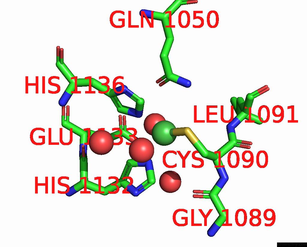
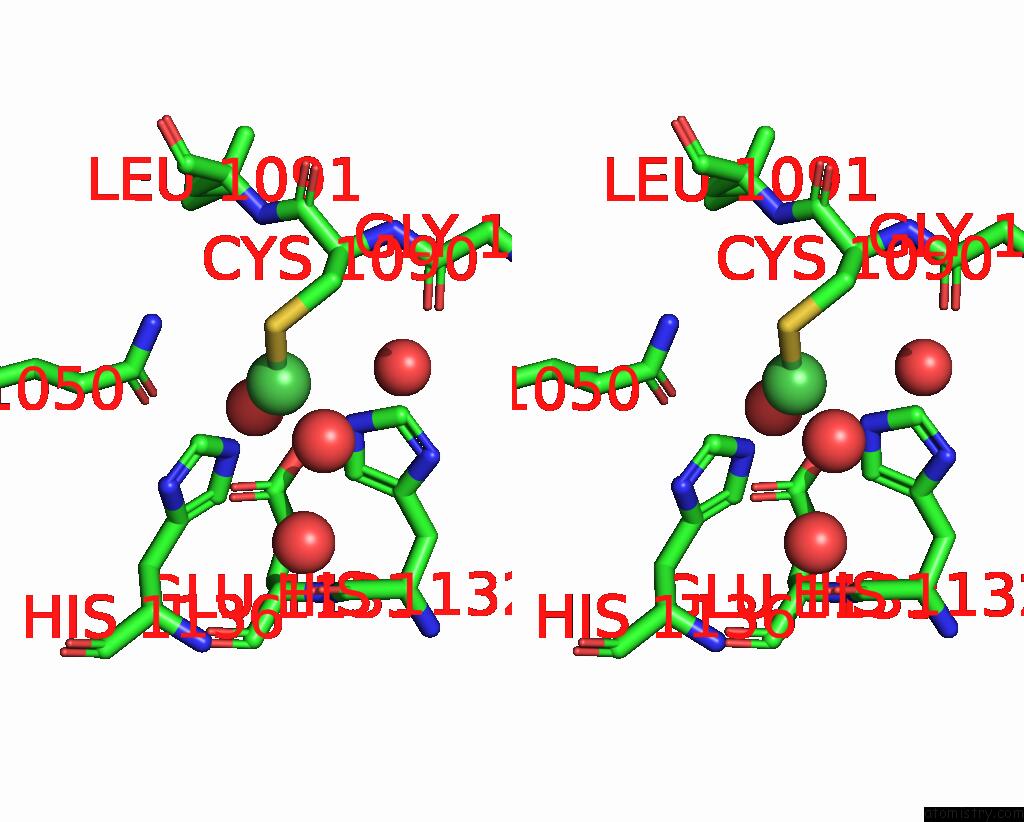
Nickel binding site 1 out of 3 in 1bs7
Go back to
Nickel binding site 1 out
of 3 in the Peptide Deformylase As NI2+ Containing Form
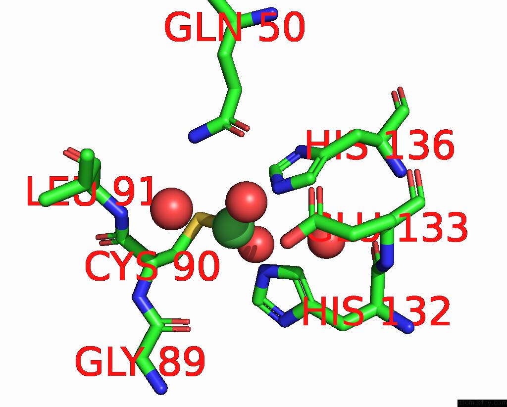
Mono view
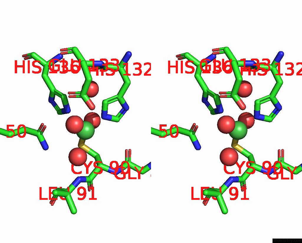
Stereo pair view
Mono view
Stereo pair view
A full contact list of Nickel with other atoms in the Ni binding
site number 1 of Peptide Deformylase As NI2+ Containing Form within 5.0Å range:
|
Nickel binding site 2 out of 3 in 1bs7
Go back to
Nickel binding site 2 out
of 3 in the Peptide Deformylase As NI2+ Containing Form
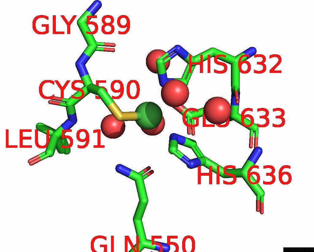
Mono view
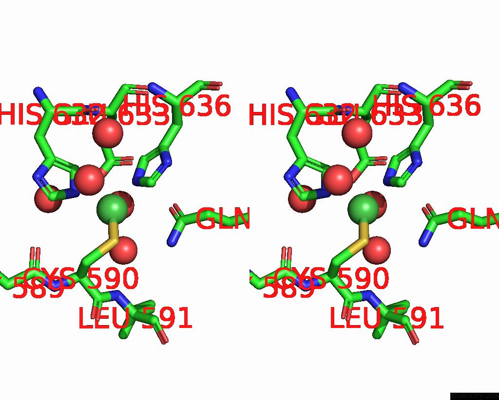
Stereo pair view
Mono view
Stereo pair view
A full contact list of Nickel with other atoms in the Ni binding
site number 2 of Peptide Deformylase As NI2+ Containing Form within 5.0Å range:
|
Nickel binding site 3 out of 3 in 1bs7
Go back to
Nickel binding site 3 out
of 3 in the Peptide Deformylase As NI2+ Containing Form
Mono view
Stereo pair view
Mono view
Stereo pair view
A full contact list of Nickel with other atoms in the Ni binding
site number 3 of Peptide Deformylase As NI2+ Containing Form within 5.0Å range:
|
Reference:
A.Becker,
I.Schlichting,
W.Kabsch,
S.Schultz,
A.F.Wagner.
Structure of Peptide Deformylase and Identification of the Substrate Binding Site. J.Biol.Chem. V. 273 11413 1998.
ISSN: ISSN 0021-9258
PubMed: 9565550
DOI: 10.1074/JBC.273.19.11413
Page generated: Wed Oct 9 14:46:28 2024
ISSN: ISSN 0021-9258
PubMed: 9565550
DOI: 10.1074/JBC.273.19.11413
Last articles
Fe in 2YXOFe in 2YRS
Fe in 2YXC
Fe in 2YNM
Fe in 2YVJ
Fe in 2YP1
Fe in 2YU2
Fe in 2YU1
Fe in 2YQB
Fe in 2YOO