Nickel »
PDB 1q0f-1sum »
1qcn »
Nickel in PDB 1qcn: Crystal Structure of Fumarylacetoacetate Hydrolase
Enzymatic activity of Crystal Structure of Fumarylacetoacetate Hydrolase
All present enzymatic activity of Crystal Structure of Fumarylacetoacetate Hydrolase:
3.7.1.2;
3.7.1.2;
Protein crystallography data
The structure of Crystal Structure of Fumarylacetoacetate Hydrolase, PDB code: 1qcn
was solved by
D.E.Timm,
H.A.Mueller,
P.Bhanumoorthy,
J.M.Harp,
G.J.Bunick,
with X-Ray Crystallography technique. A brief refinement statistics is given in the table below:
| Resolution Low / High (Å) | 22.00 / 1.90 |
| Space group | P 1 21 1 |
| Cell size a, b, c (Å), α, β, γ (°) | 64.321, 110.343, 67.533, 90.00, 102.38, 90.00 |
| R / Rfree (%) | 19.1 / 22 |
Other elements in 1qcn:
The structure of Crystal Structure of Fumarylacetoacetate Hydrolase also contains other interesting chemical elements:
| Calcium | (Ca) | 2 atoms |
Nickel Binding Sites:
The binding sites of Nickel atom in the Crystal Structure of Fumarylacetoacetate Hydrolase
(pdb code 1qcn). This binding sites where shown within
5.0 Angstroms radius around Nickel atom.
In total only one binding site of Nickel was determined in the Crystal Structure of Fumarylacetoacetate Hydrolase, PDB code: 1qcn:
In total only one binding site of Nickel was determined in the Crystal Structure of Fumarylacetoacetate Hydrolase, PDB code: 1qcn:
Nickel binding site 1 out of 1 in 1qcn
Go back to
Nickel binding site 1 out
of 1 in the Crystal Structure of Fumarylacetoacetate Hydrolase
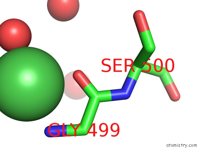
Mono view
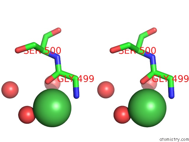
Stereo pair view
Mono view
Stereo pair view
A full contact list of Nickel with other atoms in the Ni binding
site number 1 of Crystal Structure of Fumarylacetoacetate Hydrolase within 5.0Å range:
|
Reference:
D.E.Timm,
H.A.Mueller,
P.Bhanumoorthy,
J.M.Harp,
G.J.Bunick.
Crystal Structure and Mechanism of A Carbon-Carbon Bond Hydrolase. Structure Fold.Des. V. 7 1023 1999.
ISSN: ISSN 0969-2126
PubMed: 10508789
DOI: 10.1016/S0969-2126(99)80170-1
Page generated: Wed Oct 9 16:19:03 2024
ISSN: ISSN 0969-2126
PubMed: 10508789
DOI: 10.1016/S0969-2126(99)80170-1
Last articles
Fe in 2YXOFe in 2YRS
Fe in 2YXC
Fe in 2YNM
Fe in 2YVJ
Fe in 2YP1
Fe in 2YU2
Fe in 2YU1
Fe in 2YQB
Fe in 2YOO