Nickel »
PDB 1xu1-2c0n »
2ai7 »
Nickel in PDB 2ai7: S.Pneumoniae Polypeptide Deformylase Complexed with Sb- 485345
Enzymatic activity of S.Pneumoniae Polypeptide Deformylase Complexed with Sb- 485345
All present enzymatic activity of S.Pneumoniae Polypeptide Deformylase Complexed with Sb- 485345:
3.5.1.88;
3.5.1.88;
Protein crystallography data
The structure of S.Pneumoniae Polypeptide Deformylase Complexed with Sb- 485345, PDB code: 2ai7
was solved by
K.J.Smith,
C.M.Petit,
K.Aubart,
M.Smyth,
E.Mcmanus,
J.Jones,
A.Fosberry,
C.Lewis,
M.Lonetto,
S.B.Christensen,
with X-Ray Crystallography technique. A brief refinement statistics is given in the table below:
| Resolution Low / High (Å) | 20.00 / 2.00 |
| Space group | P 43 |
| Cell size a, b, c (Å), α, β, γ (°) | 49.889, 49.889, 91.565, 90.00, 90.00, 90.00 |
| R / Rfree (%) | 22 / 26.3 |
Nickel Binding Sites:
The binding sites of Nickel atom in the S.Pneumoniae Polypeptide Deformylase Complexed with Sb- 485345
(pdb code 2ai7). This binding sites where shown within
5.0 Angstroms radius around Nickel atom.
In total only one binding site of Nickel was determined in the S.Pneumoniae Polypeptide Deformylase Complexed with Sb- 485345, PDB code: 2ai7:
In total only one binding site of Nickel was determined in the S.Pneumoniae Polypeptide Deformylase Complexed with Sb- 485345, PDB code: 2ai7:
Nickel binding site 1 out of 1 in 2ai7
Go back to
Nickel binding site 1 out
of 1 in the S.Pneumoniae Polypeptide Deformylase Complexed with Sb- 485345
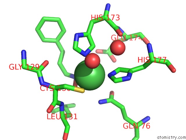
Mono view
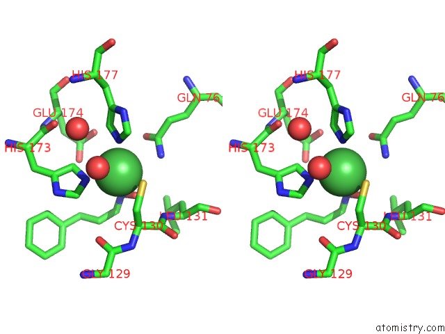
Stereo pair view
Mono view
Stereo pair view
A full contact list of Nickel with other atoms in the Ni binding
site number 1 of S.Pneumoniae Polypeptide Deformylase Complexed with Sb- 485345 within 5.0Å range:
|
Reference:
K.J.Smith,
C.M.Petit,
K.Aubart,
M.Smyth,
E.Mcmanus,
J.Jones,
A.Fosberry,
C.Lewis,
M.Lonetto,
S.B.Christensen.
Structural Variation and Inhibitor Binding in Polypeptide Deformylase From Four Different Bacterial Species Protein Sci. V. 12 349 2003.
ISSN: ISSN 0961-8368
PubMed: 12538898
DOI: 10.1110/PS.0229303
Page generated: Wed Oct 9 16:36:19 2024
ISSN: ISSN 0961-8368
PubMed: 12538898
DOI: 10.1110/PS.0229303
Last articles
Mg in 5MMJMg in 5MRA
Mg in 5MTV
Mg in 5MS0
Mg in 5MRU
Mg in 5MQJ
Mg in 5MQW
Mg in 5MQT
Mg in 5MQL
Mg in 5MQ1