Nickel »
PDB 1xu1-2c0n »
2atf »
Nickel in PDB 2atf: X-Ray Structure of Cysteine Dioxygenase Type I From Mus Musculus Mm.241056
Enzymatic activity of X-Ray Structure of Cysteine Dioxygenase Type I From Mus Musculus Mm.241056
All present enzymatic activity of X-Ray Structure of Cysteine Dioxygenase Type I From Mus Musculus Mm.241056:
1.13.11.20;
1.13.11.20;
Protein crystallography data
The structure of X-Ray Structure of Cysteine Dioxygenase Type I From Mus Musculus Mm.241056, PDB code: 2atf
was solved by
G.E.Wesenberg,
G.N.Phillips Jr.,
J.G.Mccoy,
E.Bitto,
C.A.Bingman,
S.T.M.Allard,
Center For Eukaryotic Structural Genomics (Cesg),
with X-Ray Crystallography technique. A brief refinement statistics is given in the table below:
| Resolution Low / High (Å) | 33.86 / 1.75 |
| Space group | P 43 21 2 |
| Cell size a, b, c (Å), α, β, γ (°) | 57.550, 57.550, 122.076, 90.00, 90.00, 90.00 |
| R / Rfree (%) | 17.9 / 21.6 |
Nickel Binding Sites:
The binding sites of Nickel atom in the X-Ray Structure of Cysteine Dioxygenase Type I From Mus Musculus Mm.241056
(pdb code 2atf). This binding sites where shown within
5.0 Angstroms radius around Nickel atom.
In total only one binding site of Nickel was determined in the X-Ray Structure of Cysteine Dioxygenase Type I From Mus Musculus Mm.241056, PDB code: 2atf:
In total only one binding site of Nickel was determined in the X-Ray Structure of Cysteine Dioxygenase Type I From Mus Musculus Mm.241056, PDB code: 2atf:
Nickel binding site 1 out of 1 in 2atf
Go back to
Nickel binding site 1 out
of 1 in the X-Ray Structure of Cysteine Dioxygenase Type I From Mus Musculus Mm.241056

Mono view
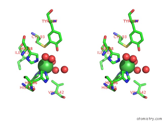
Stereo pair view
Mono view
Stereo pair view
A full contact list of Nickel with other atoms in the Ni binding
site number 1 of X-Ray Structure of Cysteine Dioxygenase Type I From Mus Musculus Mm.241056 within 5.0Å range:
|
Reference:
J.G.Mccoy,
L.J.Bailey,
E.Bitto,
C.A.Bingman,
D.J.Aceti,
B.G.Fox,
G.N.Phillips.
Structure and Mechanism of Mouse Cysteine Dioxygenase. Proc.Natl.Acad.Sci.Usa V. 103 3084 2006.
ISSN: ISSN 0027-8424
PubMed: 16492780
DOI: 10.1073/PNAS.0509262103
Page generated: Wed Oct 9 16:37:49 2024
ISSN: ISSN 0027-8424
PubMed: 16492780
DOI: 10.1073/PNAS.0509262103
Last articles
Mg in 5MMJMg in 5MRA
Mg in 5MTV
Mg in 5MS0
Mg in 5MRU
Mg in 5MQJ
Mg in 5MQW
Mg in 5MQT
Mg in 5MQL
Mg in 5MQ1