Nickel »
PDB 2c21-2gw3 »
2dro »
Nickel in PDB 2dro: Crystal Structure of Reducing-End-Xylose Releasing Exo-Oligoxylanase D263C Mutant
Enzymatic activity of Crystal Structure of Reducing-End-Xylose Releasing Exo-Oligoxylanase D263C Mutant
All present enzymatic activity of Crystal Structure of Reducing-End-Xylose Releasing Exo-Oligoxylanase D263C Mutant:
3.2.1.156;
3.2.1.156;
Protein crystallography data
The structure of Crystal Structure of Reducing-End-Xylose Releasing Exo-Oligoxylanase D263C Mutant, PDB code: 2dro
was solved by
S.Fushinobu,
M.Hidaka,
Y.Honda,
T.Wakagi,
H.Shoun,
M.Kitaoka,
with X-Ray Crystallography technique. A brief refinement statistics is given in the table below:
| Resolution Low / High (Å) | 39.07 / 1.70 |
| Space group | P 21 21 21 |
| Cell size a, b, c (Å), α, β, γ (°) | 53.242, 86.369, 87.653, 90.00, 90.00, 90.00 |
| R / Rfree (%) | 16.1 / 18.7 |
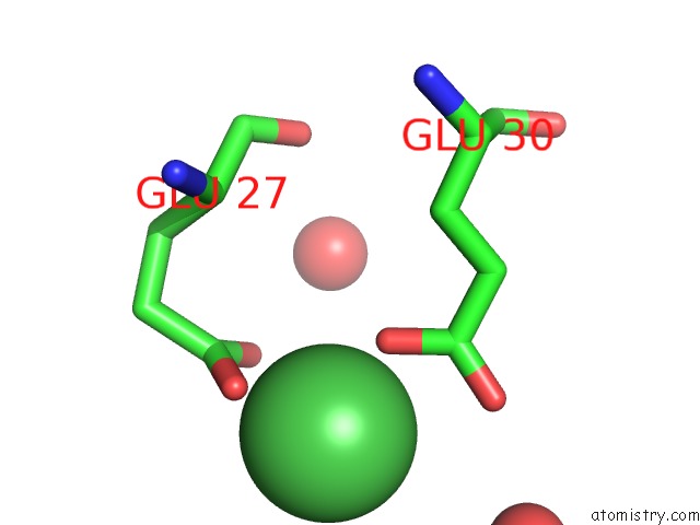
Nickel Binding Sites:
The binding sites of Nickel atom in the Crystal Structure of Reducing-End-Xylose Releasing Exo-Oligoxylanase D263C Mutant
(pdb code 2dro). This binding sites where shown within
5.0 Angstroms radius around Nickel atom.
In total only one binding site of Nickel was determined in the Crystal Structure of Reducing-End-Xylose Releasing Exo-Oligoxylanase D263C Mutant, PDB code: 2dro:
In total only one binding site of Nickel was determined in the Crystal Structure of Reducing-End-Xylose Releasing Exo-Oligoxylanase D263C Mutant, PDB code: 2dro:
Nickel binding site 1 out of 1 in 2dro
Go back to
Nickel binding site 1 out
of 1 in the Crystal Structure of Reducing-End-Xylose Releasing Exo-Oligoxylanase D263C Mutant
Mono view
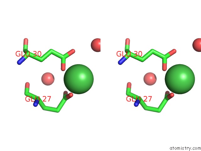
Stereo pair view
Mono view
Stereo pair view
A full contact list of Nickel with other atoms in the Ni binding
site number 1 of Crystal Structure of Reducing-End-Xylose Releasing Exo-Oligoxylanase D263C Mutant within 5.0Å range:
|
Reference:
M.Hidaka,
S.Fushinobu,
Y.Honda,
T.Wakagi,
H.Shoun,
M.Kitaoka.
Structural Explanation For the Acquisition of Glycosynthase Activity J.Biochem. 2009.
ISSN: ISSN 0021-924X
PubMed: 19819900
DOI: 10.1093/JB/MVP159
Page generated: Mon Aug 18 18:10:29 2025
ISSN: ISSN 0021-924X
PubMed: 19819900
DOI: 10.1093/JB/MVP159
Last articles
Rb in 4Z3PRb in 3HWB
Rb in 4NGJ
Rb in 4IBL
Rb in 4NGI
Rb in 4JCA
Rb in 4ILB
Rb in 3KDP
Rb in 3MGR
Rb in 461D