Nickel »
PDB 2c21-2gw3 »
2esu »
Nickel in PDB 2esu: Crystal Structure of Asn to Gln Mutant of Winged Bean Chymotrypsin Inhibitor Protein
Protein crystallography data
The structure of Crystal Structure of Asn to Gln Mutant of Winged Bean Chymotrypsin Inhibitor Protein, PDB code: 2esu
was solved by
J.K.Dattagupta,
U.Sen,
J.Dasgupta,
S.Khamrui,
with X-Ray Crystallography technique. A brief refinement statistics is given in the table below:
| Resolution Low / High (Å) | 15.00 / 1.94 |
| Space group | P 61 2 2 |
| Cell size a, b, c (Å), α, β, γ (°) | 60.900, 60.900, 213.210, 90.00, 90.00, 120.00 |
| R / Rfree (%) | 21.9 / 23.8 |
Nickel Binding Sites:
The binding sites of Nickel atom in the Crystal Structure of Asn to Gln Mutant of Winged Bean Chymotrypsin Inhibitor Protein
(pdb code 2esu). This binding sites where shown within
5.0 Angstroms radius around Nickel atom.
In total only one binding site of Nickel was determined in the Crystal Structure of Asn to Gln Mutant of Winged Bean Chymotrypsin Inhibitor Protein, PDB code: 2esu:
In total only one binding site of Nickel was determined in the Crystal Structure of Asn to Gln Mutant of Winged Bean Chymotrypsin Inhibitor Protein, PDB code: 2esu:
Nickel binding site 1 out of 1 in 2esu
Go back to
Nickel binding site 1 out
of 1 in the Crystal Structure of Asn to Gln Mutant of Winged Bean Chymotrypsin Inhibitor Protein
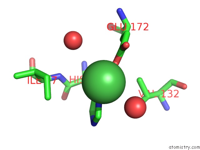
Mono view
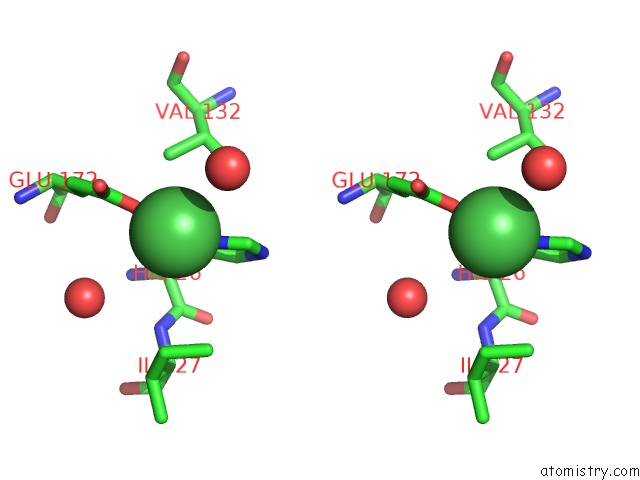
Stereo pair view
Mono view
Stereo pair view
A full contact list of Nickel with other atoms in the Ni binding
site number 1 of Crystal Structure of Asn to Gln Mutant of Winged Bean Chymotrypsin Inhibitor Protein within 5.0Å range:
|
Reference:
J.Dasgupta,
S.Khamrui,
J.K.Dattagupta,
U.Sen.
Spacer Asn Determines the Fate of Kunitz (Sti) Inhibitors, As Revealed By Structural and Biochemical Studies on Wci Mutants. Biochemistry V. 45 6783 2006.
ISSN: ISSN 0006-2960
PubMed: 16734415
DOI: 10.1021/BI060374Q
Page generated: Mon Aug 18 18:10:57 2025
ISSN: ISSN 0006-2960
PubMed: 16734415
DOI: 10.1021/BI060374Q
Last articles
Pt in 6NJWPt in 6MXO
Pt in 6HJU
Pt in 6MW5
Pt in 6LNZ
Pt in 6HJT
Pt in 6G5Y
Pt in 6GOH
Pt in 6G5V
Pt in 6GOJ