Nickel »
PDB 2gw4-2pos »
2hje »
Nickel in PDB 2hje: Crystal Structure of Vibrio Harveyi Luxq Periplasmic Domain
Enzymatic activity of Crystal Structure of Vibrio Harveyi Luxq Periplasmic Domain
All present enzymatic activity of Crystal Structure of Vibrio Harveyi Luxq Periplasmic Domain:
2.7.13.3;
2.7.13.3;
Protein crystallography data
The structure of Crystal Structure of Vibrio Harveyi Luxq Periplasmic Domain, PDB code: 2hje
was solved by
M.B.Neiditch,
R.C.Kelly,
F.M.Hughson,
with X-Ray Crystallography technique. A brief refinement statistics is given in the table below:
| Resolution Low / High (Å) | 26.10 / 1.70 |
| Space group | P 1 |
| Cell size a, b, c (Å), α, β, γ (°) | 33.446, 36.152, 49.185, 82.18, 86.14, 67.28 |
| R / Rfree (%) | 19.4 / 23.7 |
Nickel Binding Sites:
The binding sites of Nickel atom in the Crystal Structure of Vibrio Harveyi Luxq Periplasmic Domain
(pdb code 2hje). This binding sites where shown within
5.0 Angstroms radius around Nickel atom.
In total 4 binding sites of Nickel where determined in the Crystal Structure of Vibrio Harveyi Luxq Periplasmic Domain, PDB code: 2hje:
Jump to Nickel binding site number: 1; 2; 3; 4;
In total 4 binding sites of Nickel where determined in the Crystal Structure of Vibrio Harveyi Luxq Periplasmic Domain, PDB code: 2hje:
Jump to Nickel binding site number: 1; 2; 3; 4;
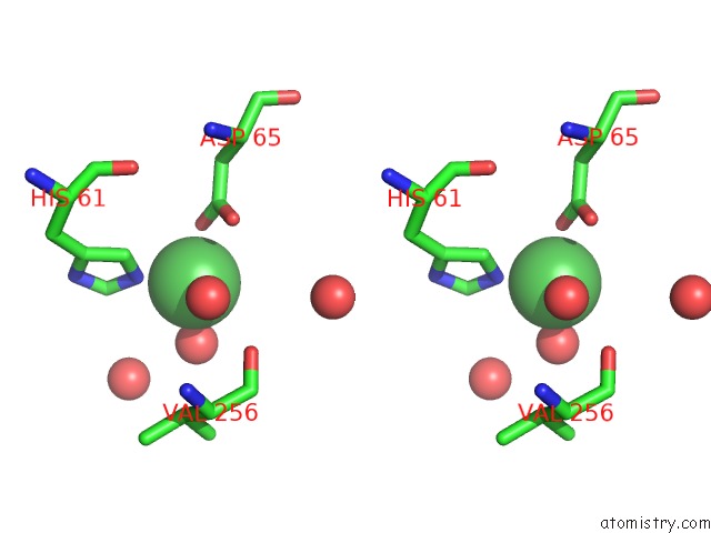
Nickel binding site 1 out of 4 in 2hje
Go back to
Nickel binding site 1 out
of 4 in the Crystal Structure of Vibrio Harveyi Luxq Periplasmic Domain

Mono view

Stereo pair view
Mono view
Stereo pair view
A full contact list of Nickel with other atoms in the Ni binding
site number 1 of Crystal Structure of Vibrio Harveyi Luxq Periplasmic Domain within 5.0Å range:
|
Nickel binding site 2 out of 4 in 2hje
Go back to
Nickel binding site 2 out
of 4 in the Crystal Structure of Vibrio Harveyi Luxq Periplasmic Domain

Mono view

Stereo pair view
Mono view
Stereo pair view
A full contact list of Nickel with other atoms in the Ni binding
site number 2 of Crystal Structure of Vibrio Harveyi Luxq Periplasmic Domain within 5.0Å range:
|
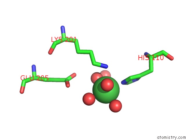
Nickel binding site 3 out of 4 in 2hje
Go back to
Nickel binding site 3 out
of 4 in the Crystal Structure of Vibrio Harveyi Luxq Periplasmic Domain

Mono view
Stereo pair view
Mono view
Stereo pair view
A full contact list of Nickel with other atoms in the Ni binding
site number 3 of Crystal Structure of Vibrio Harveyi Luxq Periplasmic Domain within 5.0Å range:
|
Nickel binding site 4 out of 4 in 2hje
Go back to
Nickel binding site 4 out
of 4 in the Crystal Structure of Vibrio Harveyi Luxq Periplasmic Domain
Mono view

Stereo pair view
Mono view
Stereo pair view
A full contact list of Nickel with other atoms in the Ni binding
site number 4 of Crystal Structure of Vibrio Harveyi Luxq Periplasmic Domain within 5.0Å range:
|
Reference:
M.B.Neiditch,
M.J.Federle,
A.J.Pompeani,
R.C.Kelly,
D.L.Swem,
P.D.Jeffrey,
B.L.Bassler,
F.M.Hughson.
Ligand-Induced Asymmetry in Histidine Sensor Kinase Complex Regulates Quorum Sensing. Cell(Cambridge,Mass.) V. 126 1095 2006.
ISSN: ISSN 0092-8674
PubMed: 16990134
DOI: 10.1016/J.CELL.2006.07.032
Page generated: Wed Oct 9 16:46:55 2024
ISSN: ISSN 0092-8674
PubMed: 16990134
DOI: 10.1016/J.CELL.2006.07.032
Last articles
Fe in 2YXOFe in 2YRS
Fe in 2YXC
Fe in 2YNM
Fe in 2YVJ
Fe in 2YP1
Fe in 2YU2
Fe in 2YU1
Fe in 2YQB
Fe in 2YOO