Nickel »
PDB 2pr0-2w3u »
2qnk »
Nickel in PDB 2qnk: Crystal Structure of Human 3-Hydroxyanthranilate 3,4-Dioxygenase
Enzymatic activity of Crystal Structure of Human 3-Hydroxyanthranilate 3,4-Dioxygenase
All present enzymatic activity of Crystal Structure of Human 3-Hydroxyanthranilate 3,4-Dioxygenase:
1.13.11.6;
1.13.11.6;
Protein crystallography data
The structure of Crystal Structure of Human 3-Hydroxyanthranilate 3,4-Dioxygenase, PDB code: 2qnk
was solved by
E.Bitto,
C.A.Bingman,
G.E.Wesenberg,
G.N.Phillips Jr.,
Center Foreukaryotic Structural Genomics (Cesg),
with X-Ray Crystallography technique. A brief refinement statistics is given in the table below:
| Resolution Low / High (Å) | 30.92 / 1.60 |
| Space group | P 21 21 21 |
| Cell size a, b, c (Å), α, β, γ (°) | 50.498, 78.227, 85.080, 90.00, 90.00, 90.00 |
| R / Rfree (%) | 15.7 / 17.6 |
Nickel Binding Sites:
The binding sites of Nickel atom in the Crystal Structure of Human 3-Hydroxyanthranilate 3,4-Dioxygenase
(pdb code 2qnk). This binding sites where shown within
5.0 Angstroms radius around Nickel atom.
In total only one binding site of Nickel was determined in the Crystal Structure of Human 3-Hydroxyanthranilate 3,4-Dioxygenase, PDB code: 2qnk:
In total only one binding site of Nickel was determined in the Crystal Structure of Human 3-Hydroxyanthranilate 3,4-Dioxygenase, PDB code: 2qnk:
Nickel binding site 1 out of 1 in 2qnk
Go back to
Nickel binding site 1 out
of 1 in the Crystal Structure of Human 3-Hydroxyanthranilate 3,4-Dioxygenase
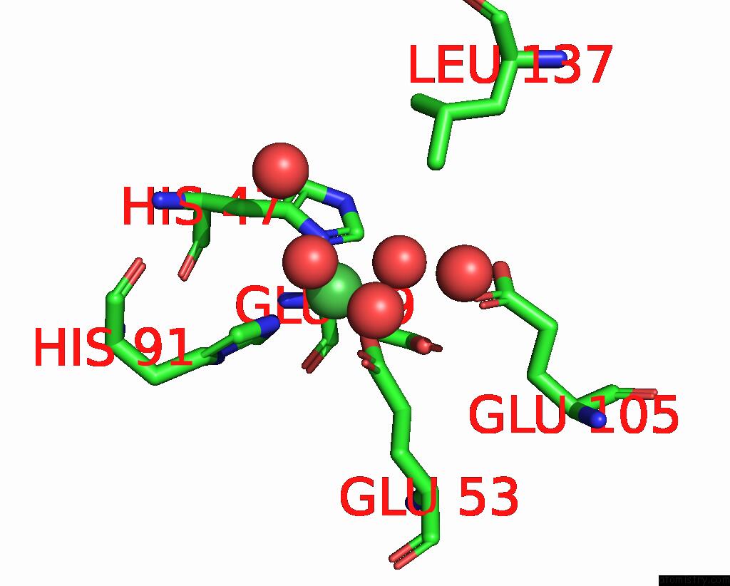
Mono view
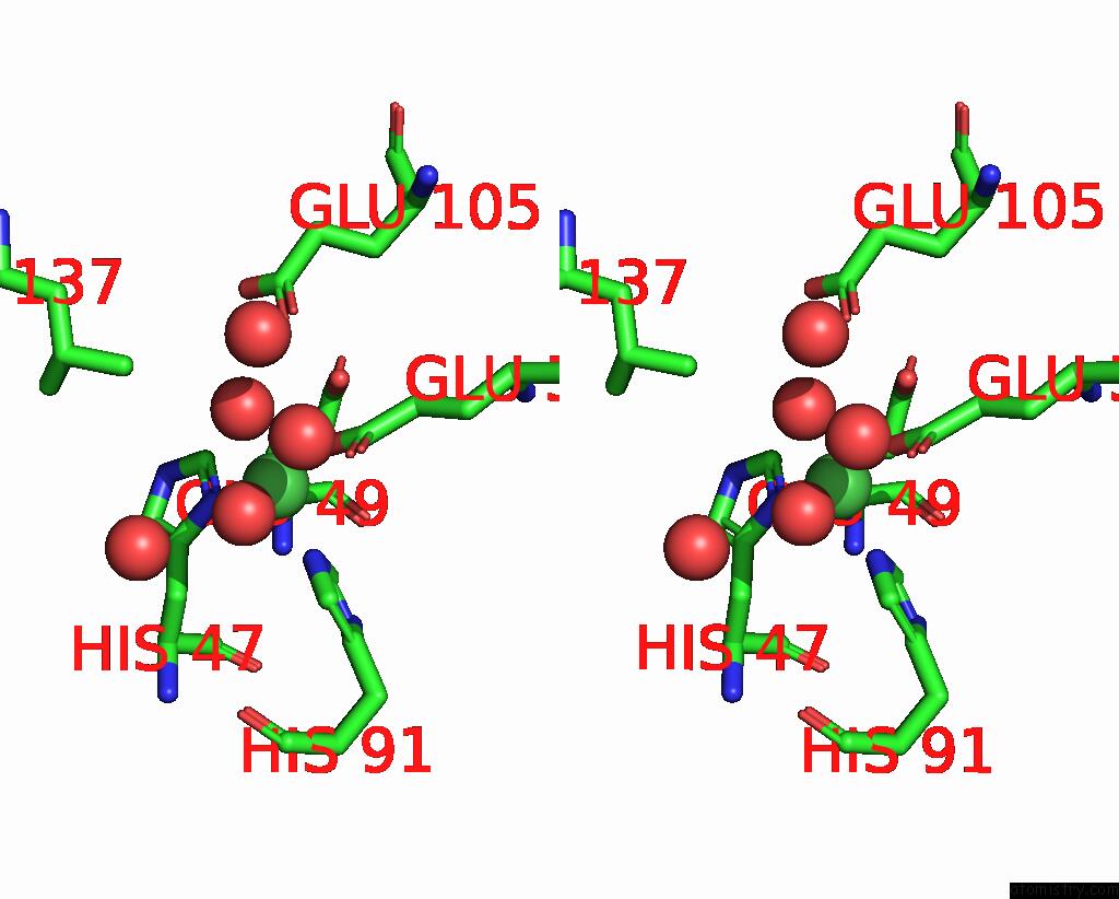
Stereo pair view
Mono view
Stereo pair view
A full contact list of Nickel with other atoms in the Ni binding
site number 1 of Crystal Structure of Human 3-Hydroxyanthranilate 3,4-Dioxygenase within 5.0Å range:
|
Reference:
E.Bitto,
C.A.Bingman,
G.E.Wesenberg,
G.N.Phillips Jr..
Crystal Structure of Human 3-Hydroxyanthranilate 3,4-Dioxygenase. To Be Published.
Page generated: Wed Oct 9 16:56:01 2024
Last articles
Na in 6ZT3Na in 6ZUK
Na in 6ZT2
Na in 6ZSN
Na in 6ZPL
Na in 6ZRA
Na in 6ZR6
Na in 6ZPQ
Na in 6ZO8
Na in 6ZO7