Nickel »
PDB 3a1f-3ds9 »
3a3v »
Nickel in PDB 3a3v: Crystal Structure of Reducing-End-Xylose Releasing Exo-Oligoxylanase Y198F Mutant
Enzymatic activity of Crystal Structure of Reducing-End-Xylose Releasing Exo-Oligoxylanase Y198F Mutant
All present enzymatic activity of Crystal Structure of Reducing-End-Xylose Releasing Exo-Oligoxylanase Y198F Mutant:
3.2.1.156;
3.2.1.156;
Protein crystallography data
The structure of Crystal Structure of Reducing-End-Xylose Releasing Exo-Oligoxylanase Y198F Mutant, PDB code: 3a3v
was solved by
M.Hidaka,
S.Fushinobu,
Y.Honda,
M.Kitaoka,
with X-Ray Crystallography technique. A brief refinement statistics is given in the table below:
| Resolution Low / High (Å) | 45.50 / 1.39 |
| Space group | P 21 21 21 |
| Cell size a, b, c (Å), α, β, γ (°) | 53.191, 86.207, 87.729, 90.00, 90.00, 90.00 |
| R / Rfree (%) | 17.7 / 19.8 |
Nickel Binding Sites:
The binding sites of Nickel atom in the Crystal Structure of Reducing-End-Xylose Releasing Exo-Oligoxylanase Y198F Mutant
(pdb code 3a3v). This binding sites where shown within
5.0 Angstroms radius around Nickel atom.
In total only one binding site of Nickel was determined in the Crystal Structure of Reducing-End-Xylose Releasing Exo-Oligoxylanase Y198F Mutant, PDB code: 3a3v:
In total only one binding site of Nickel was determined in the Crystal Structure of Reducing-End-Xylose Releasing Exo-Oligoxylanase Y198F Mutant, PDB code: 3a3v:
Nickel binding site 1 out of 1 in 3a3v
Go back to
Nickel binding site 1 out
of 1 in the Crystal Structure of Reducing-End-Xylose Releasing Exo-Oligoxylanase Y198F Mutant
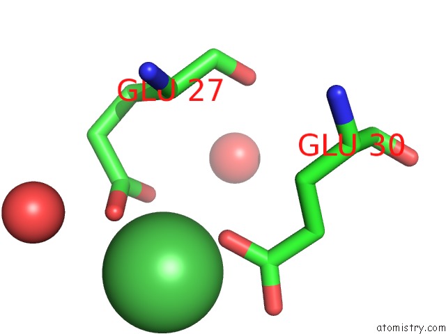
Mono view
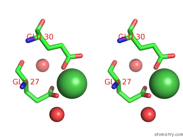
Stereo pair view
Mono view
Stereo pair view
A full contact list of Nickel with other atoms in the Ni binding
site number 1 of Crystal Structure of Reducing-End-Xylose Releasing Exo-Oligoxylanase Y198F Mutant within 5.0Å range:
|
Reference:
M.Hidaka,
S.Fushinobu,
Y.Honda,
T.Wakagi,
H.Shoun,
M.Kitaoka.
Structural Explanation For the Acquisition of Glycosynthase Activity J.Biochem. V. 147 237 2010.
ISSN: ISSN 0021-924X
PubMed: 19819900
DOI: 10.1093/JB/MVP159
Page generated: Wed Oct 9 17:10:36 2024
ISSN: ISSN 0021-924X
PubMed: 19819900
DOI: 10.1093/JB/MVP159
Last articles
Mn in 1CKNMn in 1CJT
Mn in 1CJK
Mn in 1CE8
Mn in 1CEV
Mn in 1BFR
Mn in 1C3O
Mn in 1CDK
Mn in 1C30
Mn in 1C57