Nickel »
PDB 3a1f-3ds9 »
3bq8 »
Nickel in PDB 3bq8: Crystal Structure of the E.Coli Phoq Sensor Domain
Enzymatic activity of Crystal Structure of the E.Coli Phoq Sensor Domain
All present enzymatic activity of Crystal Structure of the E.Coli Phoq Sensor Domain:
2.7.13.3;
2.7.13.3;
Protein crystallography data
The structure of Crystal Structure of the E.Coli Phoq Sensor Domain, PDB code: 3bq8
was solved by
J.Cheung,
W.A.Hendrickson,
C.D.Waldburger,
with X-Ray Crystallography technique. A brief refinement statistics is given in the table below:
| Resolution Low / High (Å) | 19.17 / 2.50 |
| Space group | P 1 21 1 |
| Cell size a, b, c (Å), α, β, γ (°) | 33.591, 113.965, 45.528, 90.00, 109.45, 90.00 |
| R / Rfree (%) | 20.3 / 30.2 |
Nickel Binding Sites:
The binding sites of Nickel atom in the Crystal Structure of the E.Coli Phoq Sensor Domain
(pdb code 3bq8). This binding sites where shown within
5.0 Angstroms radius around Nickel atom.
In total 2 binding sites of Nickel where determined in the Crystal Structure of the E.Coli Phoq Sensor Domain, PDB code: 3bq8:
Jump to Nickel binding site number: 1; 2;
In total 2 binding sites of Nickel where determined in the Crystal Structure of the E.Coli Phoq Sensor Domain, PDB code: 3bq8:
Jump to Nickel binding site number: 1; 2;
Nickel binding site 1 out of 2 in 3bq8
Go back to
Nickel binding site 1 out
of 2 in the Crystal Structure of the E.Coli Phoq Sensor Domain
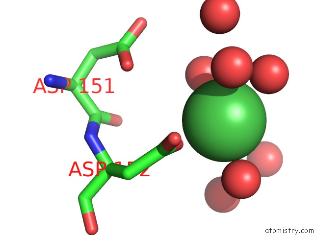
Mono view
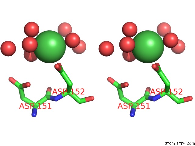
Stereo pair view
Mono view
Stereo pair view
A full contact list of Nickel with other atoms in the Ni binding
site number 1 of Crystal Structure of the E.Coli Phoq Sensor Domain within 5.0Å range:
|
Nickel binding site 2 out of 2 in 3bq8
Go back to
Nickel binding site 2 out
of 2 in the Crystal Structure of the E.Coli Phoq Sensor Domain
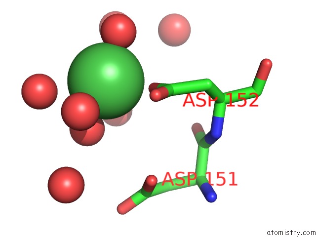
Mono view

Stereo pair view
Mono view
Stereo pair view
A full contact list of Nickel with other atoms in the Ni binding
site number 2 of Crystal Structure of the E.Coli Phoq Sensor Domain within 5.0Å range:
|
Reference:
J.Cheung,
C.A.Bingman,
M.Reyngold,
W.A.Hendrickson,
C.D.Waldburger.
Crystal Structure of A Functional Dimer of the Phoq Sensor Domain. J.Biol.Chem. V. 283 13762 2008.
ISSN: ISSN 0021-9258
PubMed: 18348979
DOI: 10.1074/JBC.M710592200
Page generated: Wed Oct 9 17:12:43 2024
ISSN: ISSN 0021-9258
PubMed: 18348979
DOI: 10.1074/JBC.M710592200
Last articles
Mn in 1F5AMn in 1F3W
Mn in 1EQZ
Mn in 1F1V
Mn in 1F1U
Mn in 1F1R
Mn in 1EO4
Mn in 1EN6
Mn in 1EQJ
Mn in 1EN5