Nickel »
PDB 3a1f-3ds9 »
3cu2 »
Nickel in PDB 3cu2: Crystal Structure of Ribulose-5-Phosphate 3-Epimerase (YP_718263.1) From Haemophilus Somnus 129PT at 1.91 A Resolution
Enzymatic activity of Crystal Structure of Ribulose-5-Phosphate 3-Epimerase (YP_718263.1) From Haemophilus Somnus 129PT at 1.91 A Resolution
All present enzymatic activity of Crystal Structure of Ribulose-5-Phosphate 3-Epimerase (YP_718263.1) From Haemophilus Somnus 129PT at 1.91 A Resolution:
5.1.3.1;
5.1.3.1;
Protein crystallography data
The structure of Crystal Structure of Ribulose-5-Phosphate 3-Epimerase (YP_718263.1) From Haemophilus Somnus 129PT at 1.91 A Resolution, PDB code: 3cu2
was solved by
Joint Center For Structural Genomics (Jcsg),
with X-Ray Crystallography technique. A brief refinement statistics is given in the table below:
| Resolution Low / High (Å) | 28.92 / 1.91 |
| Space group | P 31 2 1 |
| Cell size a, b, c (Å), α, β, γ (°) | 85.010, 85.010, 140.160, 90.00, 90.00, 120.00 |
| R / Rfree (%) | 16.6 / 20.5 |
Other elements in 3cu2:
The structure of Crystal Structure of Ribulose-5-Phosphate 3-Epimerase (YP_718263.1) From Haemophilus Somnus 129PT at 1.91 A Resolution also contains other interesting chemical elements:
| Calcium | (Ca) | 2 atoms |
Nickel Binding Sites:
The binding sites of Nickel atom in the Crystal Structure of Ribulose-5-Phosphate 3-Epimerase (YP_718263.1) From Haemophilus Somnus 129PT at 1.91 A Resolution
(pdb code 3cu2). This binding sites where shown within
5.0 Angstroms radius around Nickel atom.
In total 2 binding sites of Nickel where determined in the Crystal Structure of Ribulose-5-Phosphate 3-Epimerase (YP_718263.1) From Haemophilus Somnus 129PT at 1.91 A Resolution, PDB code: 3cu2:
Jump to Nickel binding site number: 1; 2;
In total 2 binding sites of Nickel where determined in the Crystal Structure of Ribulose-5-Phosphate 3-Epimerase (YP_718263.1) From Haemophilus Somnus 129PT at 1.91 A Resolution, PDB code: 3cu2:
Jump to Nickel binding site number: 1; 2;
Nickel binding site 1 out of 2 in 3cu2
Go back to
Nickel binding site 1 out
of 2 in the Crystal Structure of Ribulose-5-Phosphate 3-Epimerase (YP_718263.1) From Haemophilus Somnus 129PT at 1.91 A Resolution
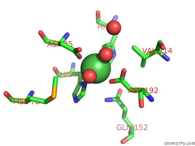
Mono view
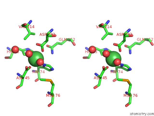
Stereo pair view
Mono view
Stereo pair view
A full contact list of Nickel with other atoms in the Ni binding
site number 1 of Crystal Structure of Ribulose-5-Phosphate 3-Epimerase (YP_718263.1) From Haemophilus Somnus 129PT at 1.91 A Resolution within 5.0Å range:
|
Nickel binding site 2 out of 2 in 3cu2
Go back to
Nickel binding site 2 out
of 2 in the Crystal Structure of Ribulose-5-Phosphate 3-Epimerase (YP_718263.1) From Haemophilus Somnus 129PT at 1.91 A Resolution
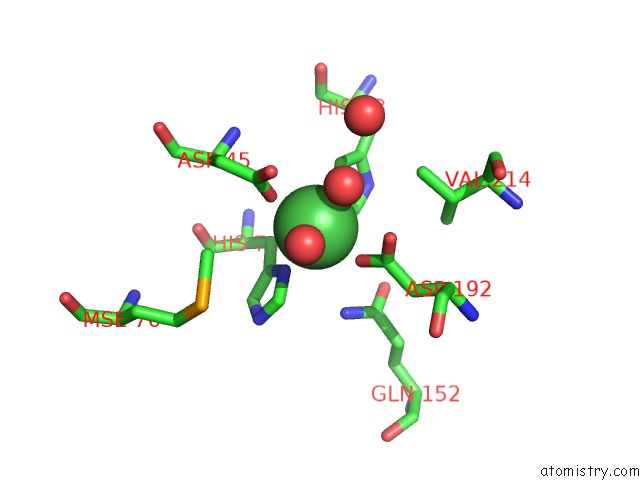
Mono view
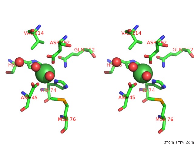
Stereo pair view
Mono view
Stereo pair view
A full contact list of Nickel with other atoms in the Ni binding
site number 2 of Crystal Structure of Ribulose-5-Phosphate 3-Epimerase (YP_718263.1) From Haemophilus Somnus 129PT at 1.91 A Resolution within 5.0Å range:
|
Reference:
Joint Center For Structural Genomics (Jcsg),
Joint Center For Structural Genomics (Jcsg).
N/A N/A.
Page generated: Wed Oct 9 17:14:04 2024
Last articles
Mg in 4LFGMg in 4LFV
Mg in 4LFA
Mg in 4LFE
Mg in 4LF8
Mg in 4LF9
Mg in 4LF7
Mg in 4LF6
Mg in 4LF4
Mg in 4LF5