Nickel »
PDB 3dse-3hy3 »
3e3u »
Nickel in PDB 3e3u: Crystal Structure of Mycobacterium Tuberculosis Peptide Deformylase in Complex with Inhibitor
Enzymatic activity of Crystal Structure of Mycobacterium Tuberculosis Peptide Deformylase in Complex with Inhibitor
All present enzymatic activity of Crystal Structure of Mycobacterium Tuberculosis Peptide Deformylase in Complex with Inhibitor:
3.5.1.88;
3.5.1.88;
Protein crystallography data
The structure of Crystal Structure of Mycobacterium Tuberculosis Peptide Deformylase in Complex with Inhibitor, PDB code: 3e3u
was solved by
W.Meng,
M.Xu,
S.Pan,
J.Koehn,
with X-Ray Crystallography technique. A brief refinement statistics is given in the table below:
| Resolution Low / High (Å) | 50.32 / 1.56 |
| Space group | C 1 2 1 |
| Cell size a, b, c (Å), α, β, γ (°) | 100.870, 54.765, 40.215, 90.00, 94.39, 90.00 |
| R / Rfree (%) | 17.1 / 20.3 |
Nickel Binding Sites:
The binding sites of Nickel atom in the Crystal Structure of Mycobacterium Tuberculosis Peptide Deformylase in Complex with Inhibitor
(pdb code 3e3u). This binding sites where shown within
5.0 Angstroms radius around Nickel atom.
In total only one binding site of Nickel was determined in the Crystal Structure of Mycobacterium Tuberculosis Peptide Deformylase in Complex with Inhibitor, PDB code: 3e3u:
In total only one binding site of Nickel was determined in the Crystal Structure of Mycobacterium Tuberculosis Peptide Deformylase in Complex with Inhibitor, PDB code: 3e3u:
Nickel binding site 1 out of 1 in 3e3u
Go back to
Nickel binding site 1 out
of 1 in the Crystal Structure of Mycobacterium Tuberculosis Peptide Deformylase in Complex with Inhibitor
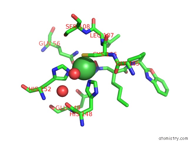
Mono view
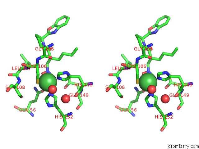
Stereo pair view
Mono view
Stereo pair view
A full contact list of Nickel with other atoms in the Ni binding
site number 1 of Crystal Structure of Mycobacterium Tuberculosis Peptide Deformylase in Complex with Inhibitor within 5.0Å range:
|
Reference:
A.Pichota,
J.Duraiswamy,
Z.Yin,
T.H.Keller,
J.Alam,
S.Liung,
G.Lee,
M.Ding,
G.Wang,
W.L.Chan,
M.Schreiber,
I.Ma,
D.Beer,
X.Ngew,
K.Mukherjee,
M.Nanjundappa,
J.W.Teo,
P.Thayalan,
A.Yap,
T.Dick,
W.Meng,
M.Xu,
J.Koehn,
S.H.Pan,
K.Clark,
X.Xie,
C.Shoen,
M.Cynamon.
Peptide Deformylase Inhibitors of Mycobacterium Tuberculosis: Synthesis, Structural Investigations, and Biological Results. Bioorg.Med.Chem.Lett. V. 18 6568 2008.
ISSN: ISSN 0960-894X
PubMed: 19008098
DOI: 10.1016/J.BMCL.2008.10.040
Page generated: Wed Oct 9 17:16:36 2024
ISSN: ISSN 0960-894X
PubMed: 19008098
DOI: 10.1016/J.BMCL.2008.10.040
Last articles
Mg in 5L5WMg in 5L5Y
Mg in 5L5X
Mg in 5L5V
Mg in 5L5U
Mg in 5L5T
Mg in 5L5S
Mg in 5L5R
Mg in 5L5P
Mg in 5L5Q