Nickel »
PDB 3dse-3hy3 »
3egj »
Nickel in PDB 3egj: N-Acetylglucosamine-6-Phosphate Deacetylase From Vibrio Cholerae.
Enzymatic activity of N-Acetylglucosamine-6-Phosphate Deacetylase From Vibrio Cholerae.
All present enzymatic activity of N-Acetylglucosamine-6-Phosphate Deacetylase From Vibrio Cholerae.:
3.5.1.25;
3.5.1.25;
Protein crystallography data
The structure of N-Acetylglucosamine-6-Phosphate Deacetylase From Vibrio Cholerae., PDB code: 3egj
was solved by
J.Osipiuk,
N.Maltseva,
J.Stam,
W.F.Anderson,
A.Joachimiak,
Center Forstructural Genomics Of Infectious Diseases (Csgid),
with X-Ray Crystallography technique. A brief refinement statistics is given in the table below:
| Resolution Low / High (Å) | 47.20 / 2.90 |
| Space group | P 43 2 2 |
| Cell size a, b, c (Å), α, β, γ (°) | 77.001, 77.001, 282.549, 90.00, 90.00, 90.00 |
| R / Rfree (%) | 18.9 / 24.7 |
Nickel Binding Sites:
The binding sites of Nickel atom in the N-Acetylglucosamine-6-Phosphate Deacetylase From Vibrio Cholerae.
(pdb code 3egj). This binding sites where shown within
5.0 Angstroms radius around Nickel atom.
In total 2 binding sites of Nickel where determined in the N-Acetylglucosamine-6-Phosphate Deacetylase From Vibrio Cholerae., PDB code: 3egj:
Jump to Nickel binding site number: 1; 2;
In total 2 binding sites of Nickel where determined in the N-Acetylglucosamine-6-Phosphate Deacetylase From Vibrio Cholerae., PDB code: 3egj:
Jump to Nickel binding site number: 1; 2;
Nickel binding site 1 out of 2 in 3egj
Go back to
Nickel binding site 1 out
of 2 in the N-Acetylglucosamine-6-Phosphate Deacetylase From Vibrio Cholerae.
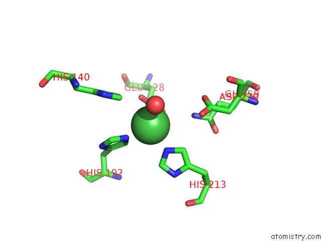
Mono view
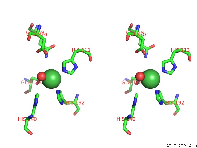
Stereo pair view
Mono view
Stereo pair view
A full contact list of Nickel with other atoms in the Ni binding
site number 1 of N-Acetylglucosamine-6-Phosphate Deacetylase From Vibrio Cholerae. within 5.0Å range:
|
Nickel binding site 2 out of 2 in 3egj
Go back to
Nickel binding site 2 out
of 2 in the N-Acetylglucosamine-6-Phosphate Deacetylase From Vibrio Cholerae.
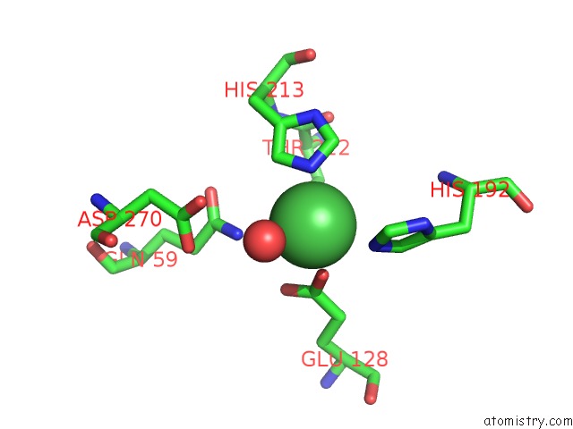
Mono view
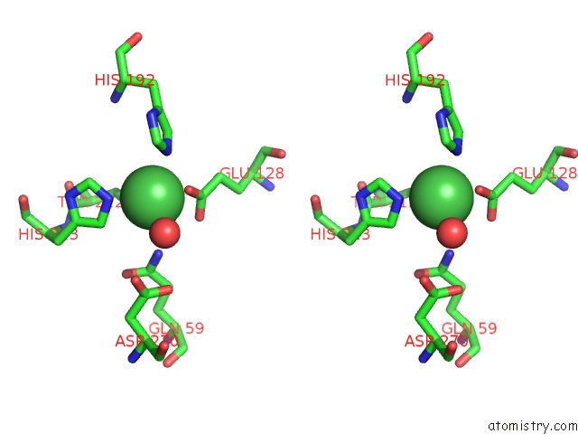
Stereo pair view
Mono view
Stereo pair view
A full contact list of Nickel with other atoms in the Ni binding
site number 2 of N-Acetylglucosamine-6-Phosphate Deacetylase From Vibrio Cholerae. within 5.0Å range:
|
Reference:
J.Osipiuk,
N.Maltseva,
J.Stam,
W.F.Anderson,
A.Joachimiak.
X-Ray Crystal Structure of N-Acetylglucosamine-6-Phosphate Deacetylase From Vibrio Cholerae. To Be Published.
Page generated: Wed Oct 9 17:16:36 2024
Last articles
Mg in 5DNNMg in 5DNM
Mg in 5DNB
Mg in 5DN6
Mg in 5DMY
Mg in 5DN3
Mg in 5DMZ
Mg in 5DMP
Mg in 5DKI
Mg in 5DKJ