Nickel »
PDB 3dse-3hy3 »
3gor »
Nickel in PDB 3gor: Crystal Structure of Putative Metal-Dependent Hydrolase APC36150
Protein crystallography data
The structure of Crystal Structure of Putative Metal-Dependent Hydrolase APC36150, PDB code: 3gor
was solved by
D.R.Cooper,
K.Grelewska,
Z.S.Derewenda,
Integrated Center For Structureand Function Innovation (Isfi),
with X-Ray Crystallography technique. A brief refinement statistics is given in the table below:
| Resolution Low / High (Å) | 35.62 / 2.51 |
| Space group | P 21 21 21 |
| Cell size a, b, c (Å), α, β, γ (°) | 68.477, 71.470, 123.256, 90.00, 90.00, 90.00 |
| R / Rfree (%) | 18.6 / 24.7 |
Nickel Binding Sites:
The binding sites of Nickel atom in the Crystal Structure of Putative Metal-Dependent Hydrolase APC36150
(pdb code 3gor). This binding sites where shown within
5.0 Angstroms radius around Nickel atom.
In total 4 binding sites of Nickel where determined in the Crystal Structure of Putative Metal-Dependent Hydrolase APC36150, PDB code: 3gor:
Jump to Nickel binding site number: 1; 2; 3; 4;
In total 4 binding sites of Nickel where determined in the Crystal Structure of Putative Metal-Dependent Hydrolase APC36150, PDB code: 3gor:
Jump to Nickel binding site number: 1; 2; 3; 4;
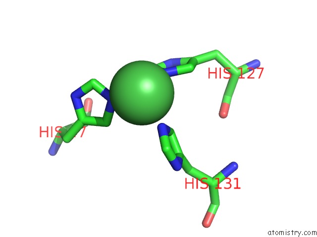
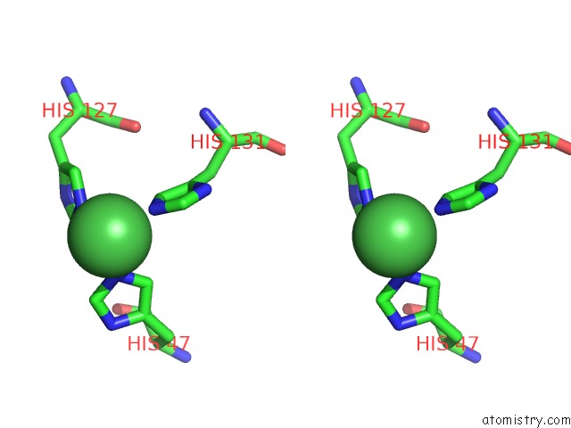
Nickel binding site 1 out of 4 in 3gor
Go back to
Nickel binding site 1 out
of 4 in the Crystal Structure of Putative Metal-Dependent Hydrolase APC36150
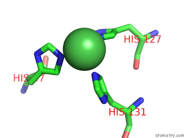
Mono view
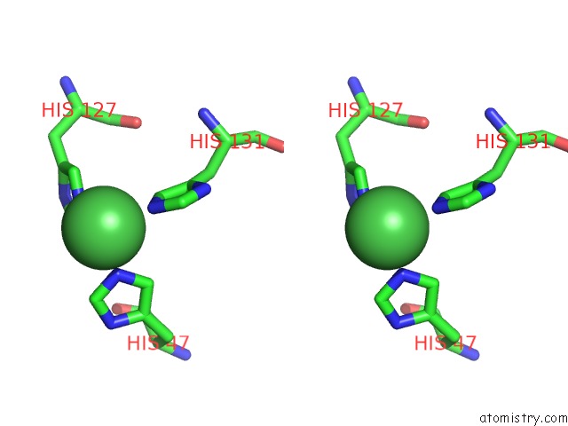
Stereo pair view
Mono view
Stereo pair view
A full contact list of Nickel with other atoms in the Ni binding
site number 1 of Crystal Structure of Putative Metal-Dependent Hydrolase APC36150 within 5.0Å range:
|
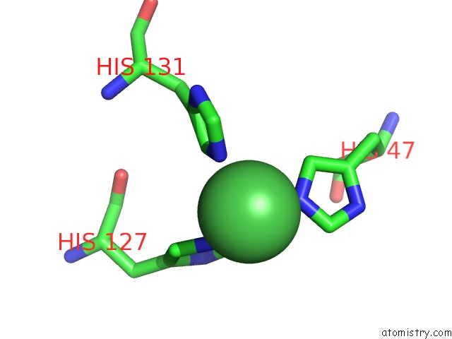
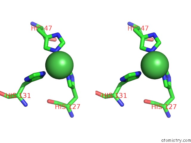
Nickel binding site 2 out of 4 in 3gor
Go back to
Nickel binding site 2 out
of 4 in the Crystal Structure of Putative Metal-Dependent Hydrolase APC36150
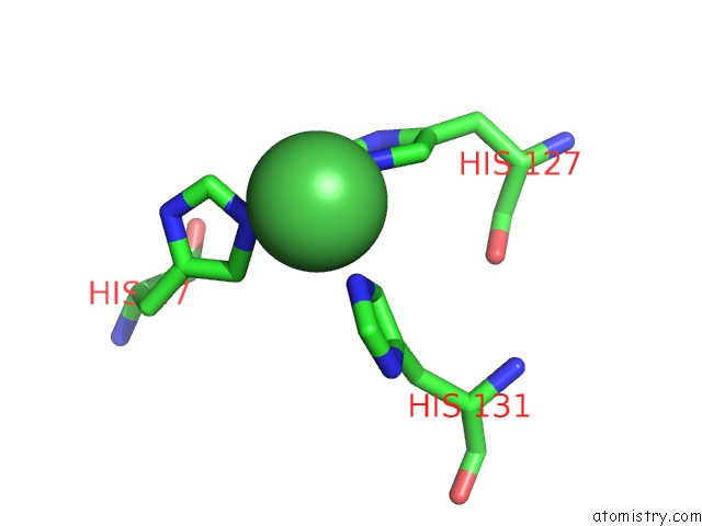
Mono view
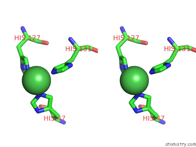
Stereo pair view
Mono view
Stereo pair view
A full contact list of Nickel with other atoms in the Ni binding
site number 2 of Crystal Structure of Putative Metal-Dependent Hydrolase APC36150 within 5.0Å range:
|
Nickel binding site 3 out of 4 in 3gor
Go back to
Nickel binding site 3 out
of 4 in the Crystal Structure of Putative Metal-Dependent Hydrolase APC36150
Mono view
Stereo pair view
Mono view
Stereo pair view
A full contact list of Nickel with other atoms in the Ni binding
site number 3 of Crystal Structure of Putative Metal-Dependent Hydrolase APC36150 within 5.0Å range:
|
Nickel binding site 4 out of 4 in 3gor
Go back to
Nickel binding site 4 out
of 4 in the Crystal Structure of Putative Metal-Dependent Hydrolase APC36150
Mono view
Stereo pair view
Mono view
Stereo pair view
A full contact list of Nickel with other atoms in the Ni binding
site number 4 of Crystal Structure of Putative Metal-Dependent Hydrolase APC36150 within 5.0Å range:
|
Reference:
D.R.Cooper,
K.Grelewska,
C.Y.Kim,
A.Joachimiak,
Z.S.Derewenda.
The Structure of Dinb From Geobacillus Stearothermophilus: A Representative of A Unique Four-Helix-Bundle Superfamily. Acta Crystallogr.,Sect.F V. 66 219 2010.
ISSN: ESSN 1744-3091
PubMed: 20208147
DOI: 10.1107/S1744309109053913
Page generated: Wed Oct 9 17:19:05 2024
ISSN: ESSN 1744-3091
PubMed: 20208147
DOI: 10.1107/S1744309109053913
Last articles
Mg in 5L5WMg in 5L5Y
Mg in 5L5X
Mg in 5L5V
Mg in 5L5U
Mg in 5L5T
Mg in 5L5S
Mg in 5L5R
Mg in 5L5P
Mg in 5L5Q