Nickel »
PDB 3hy4-3kvb »
3iar »
Nickel in PDB 3iar: The Crystal Structure of Human Adenosine Deaminase
Enzymatic activity of The Crystal Structure of Human Adenosine Deaminase
All present enzymatic activity of The Crystal Structure of Human Adenosine Deaminase:
3.5.4.4;
3.5.4.4;
Protein crystallography data
The structure of The Crystal Structure of Human Adenosine Deaminase, PDB code: 3iar
was solved by
E.Ugochukwu,
Y.Zhang,
E.Hapka,
W.W.Yue,
J.E.Bray,
J.Muniz,
N.Burgess-Brown,
A.Chaikuad,
F.Von Delft,
C.Bountra,
C.H.Arrowsmith,
J.Weigelt,
A.Edwards,
K.L.Kavanagh,
U.Oppermann,
Structural Genomics Consortium (Sgc),
with X-Ray Crystallography technique. A brief refinement statistics is given in the table below:
| Resolution Low / High (Å) | 29.70 / 1.52 |
| Space group | P 21 21 21 |
| Cell size a, b, c (Å), α, β, γ (°) | 61.070, 73.510, 76.660, 90.00, 90.00, 90.00 |
| R / Rfree (%) | 15.3 / 18.5 |
Nickel Binding Sites:
The binding sites of Nickel atom in the The Crystal Structure of Human Adenosine Deaminase
(pdb code 3iar). This binding sites where shown within
5.0 Angstroms radius around Nickel atom.
In total only one binding site of Nickel was determined in the The Crystal Structure of Human Adenosine Deaminase, PDB code: 3iar:
In total only one binding site of Nickel was determined in the The Crystal Structure of Human Adenosine Deaminase, PDB code: 3iar:
Nickel binding site 1 out of 1 in 3iar
Go back to
Nickel binding site 1 out
of 1 in the The Crystal Structure of Human Adenosine Deaminase
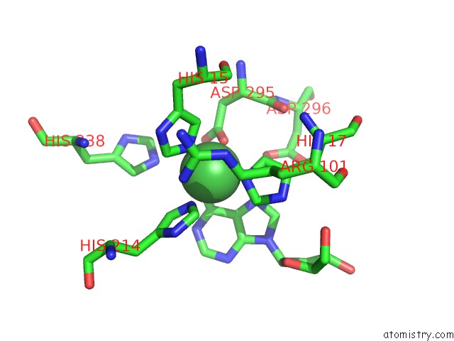
Mono view
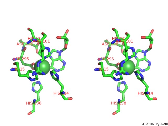
Stereo pair view
Mono view
Stereo pair view
A full contact list of Nickel with other atoms in the Ni binding
site number 1 of The Crystal Structure of Human Adenosine Deaminase within 5.0Å range:
|
Reference:
E.Ugochukwu,
Y.Zhang,
E.Hapka,
W.W.Yue,
J.E.Bray,
J.Muniz,
N.Burgess-Brown,
A.Chaikuad,
K.L.Kavanagh,
U.Oppermann.
The Crystal Structure of Human Adenosine Deaminase To Be Published.
Page generated: Wed Oct 9 17:23:22 2024
Last articles
Mg in 6DUHMg in 6DUG
Mg in 6DUF
Mg in 6DUE
Mg in 6DTQ
Mg in 6DU3
Mg in 6DU2
Mg in 6DTA
Mg in 6DTW
Mg in 6DPU