Nickel »
PDB 3l1m-3n6n »
3l1m »
Nickel in PDB 3l1m: Crystal Structure of A Ni-Directed Dimer of Cytochrome CB562 with A Quinolate-Histidine Hybrid Coordination Motif
Protein crystallography data
The structure of Crystal Structure of A Ni-Directed Dimer of Cytochrome CB562 with A Quinolate-Histidine Hybrid Coordination Motif, PDB code: 3l1m
was solved by
R.J.Radford,
F.A.Tezcan,
with X-Ray Crystallography technique. A brief refinement statistics is given in the table below:
| Resolution Low / High (Å) | 50.00 / 2.30 |
| Space group | P 21 21 2 |
| Cell size a, b, c (Å), α, β, γ (°) | 88.440, 34.511, 46.896, 90.00, 90.00, 90.00 |
| R / Rfree (%) | 26.6 / 31.4 |
Other elements in 3l1m:
The structure of Crystal Structure of A Ni-Directed Dimer of Cytochrome CB562 with A Quinolate-Histidine Hybrid Coordination Motif also contains other interesting chemical elements:
| Iron | (Fe) | 1 atom |
Nickel Binding Sites:
The binding sites of Nickel atom in the Crystal Structure of A Ni-Directed Dimer of Cytochrome CB562 with A Quinolate-Histidine Hybrid Coordination Motif
(pdb code 3l1m). This binding sites where shown within
5.0 Angstroms radius around Nickel atom.
In total only one binding site of Nickel was determined in the Crystal Structure of A Ni-Directed Dimer of Cytochrome CB562 with A Quinolate-Histidine Hybrid Coordination Motif, PDB code: 3l1m:
In total only one binding site of Nickel was determined in the Crystal Structure of A Ni-Directed Dimer of Cytochrome CB562 with A Quinolate-Histidine Hybrid Coordination Motif, PDB code: 3l1m:
Nickel binding site 1 out of 1 in 3l1m
Go back to
Nickel binding site 1 out
of 1 in the Crystal Structure of A Ni-Directed Dimer of Cytochrome CB562 with A Quinolate-Histidine Hybrid Coordination Motif
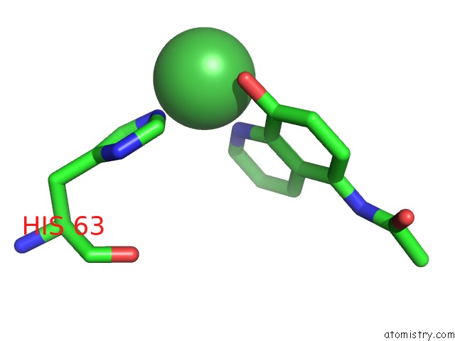
Mono view
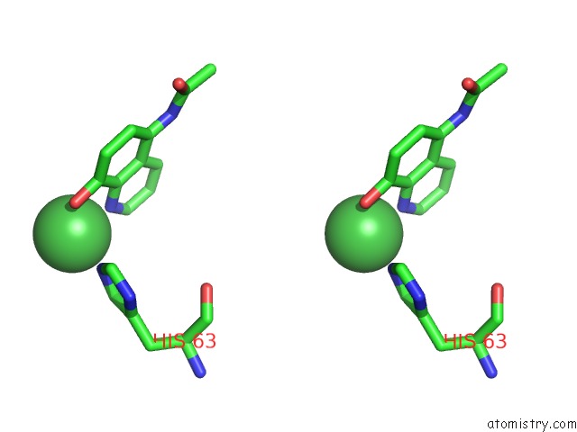
Stereo pair view
Mono view
Stereo pair view
A full contact list of Nickel with other atoms in the Ni binding
site number 1 of Crystal Structure of A Ni-Directed Dimer of Cytochrome CB562 with A Quinolate-Histidine Hybrid Coordination Motif within 5.0Å range:
|
Reference:
R.J.Radford,
P.C.Nguyen,
T.B.Ditri,
J.S.Figueroa,
F.A.Tezcan.
Controlled Protein Dimerization Through Hybrid Coordination Motifs. Inorg.Chem. V. 49 4362 2010.
ISSN: ISSN 0020-1669
PubMed: 20377257
DOI: 10.1021/IC100534Y
Page generated: Wed Oct 9 17:29:41 2024
ISSN: ISSN 0020-1669
PubMed: 20377257
DOI: 10.1021/IC100534Y
Last articles
Na in 8ALMNa in 8ALH
Na in 8AHY
Na in 8AJ3
Na in 8AGQ
Na in 8AI7
Na in 8AFV
Na in 8AHQ
Na in 8AHO
Na in 8AEB